González Estévez N1 , Priya S2, Ashwath ML 2, Ashwath RC 1.
1Stead Family Department of Pediatrics, 2University of Iowa Hospitals and Clinics, University of Iowa Health Care
Clinical history:
The patient is a 27-year-old male status post complete surgical repair of supra-cardiac total anomalous pulmonary venous return (TAPVR) in infancy. He was followed intermittently until 15 years old, with no concerning imaging findings, and was essentially asymptomatic. At 23 and 27 years of age, he presented to the emergency department with atrial fibrillation and atrial flutter, respectively, requiring cardioversion. He had a transthoracic echocardiogram (TTE) performed which showed concerning findings of a mildly dilated inferior vena cava (IVC) and hepatic veins with exaggerated reversal of flow (Movie 1, Figure 1), dilated right ventricle (RV), abnormal venous flow pattern in the right atrium (Movie 1), and a dilated venous confluence (Movie 2, Figure 2)
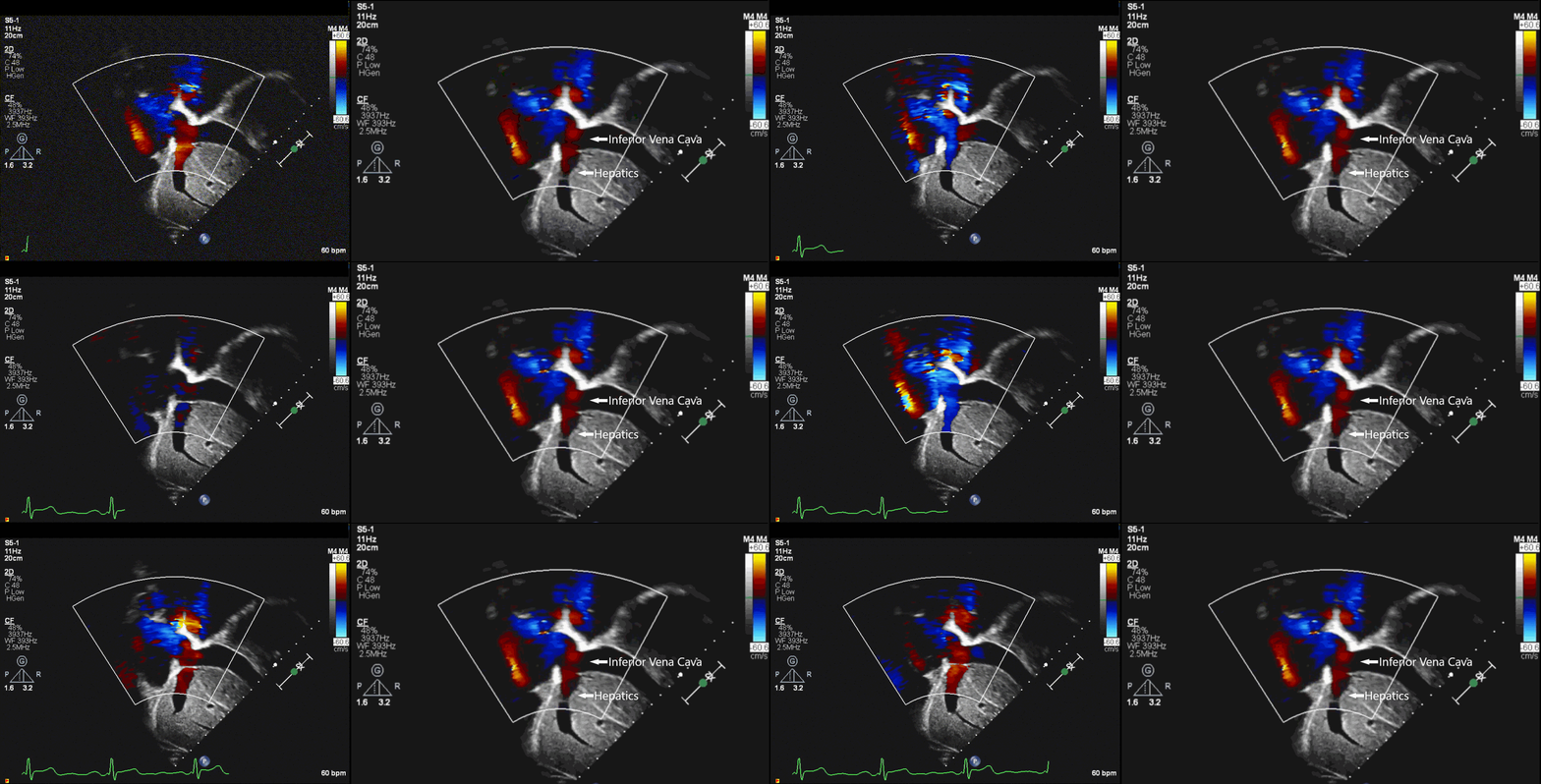
Movie 1: Bicaval view showing a dilated IVC and hepatics with flow reversal. Turbulent venous flow into the right atrium is also evident.
Figure 1. Bicaval view showing a dilated IVC and hepatics with flow reversal.
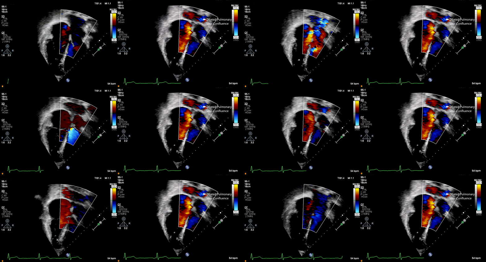
Movie 2: Color flow mapping in apical four chamber view showing the dilated pulmonary vein confluence.
Figure 2. Color flow mapping in apical four chamber view showing the dilated pulmonary vein confluence.
CMR Findings:
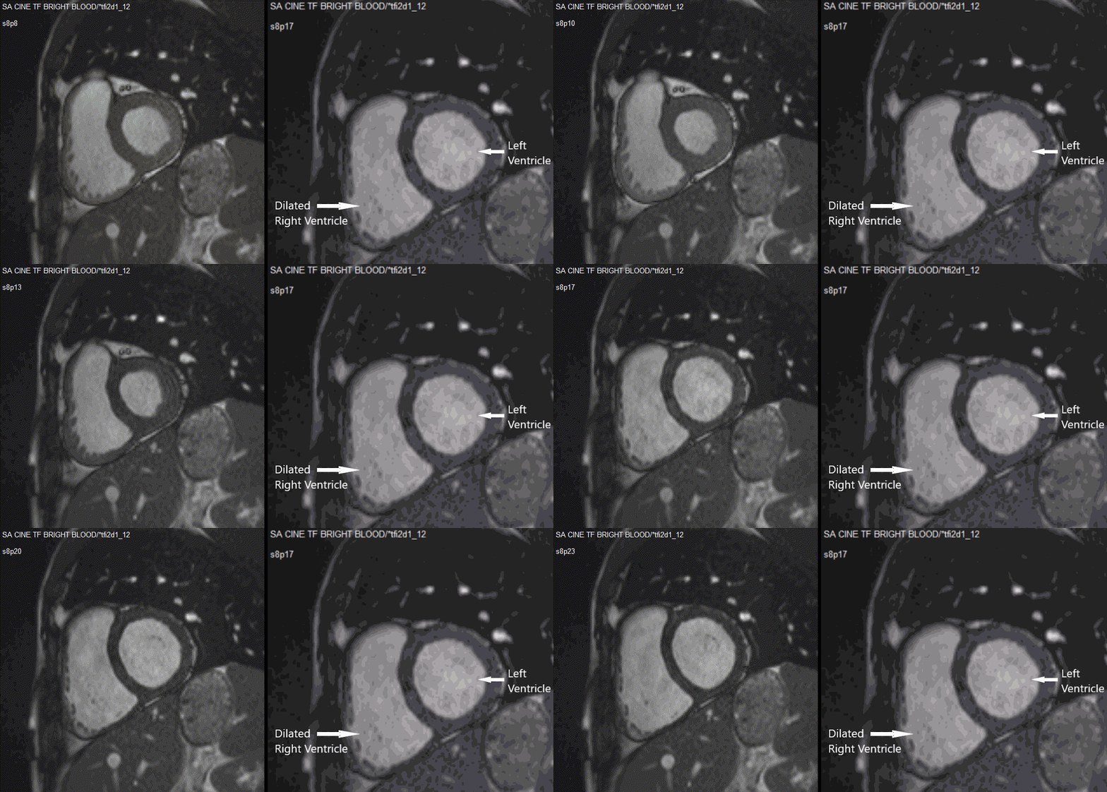
A cardiac magnetic resonance imaging (CMR) study was performed to assess pulmonary veins, venous confluence, and unexplained right ventricular dilation (Movie 3, Figure 3).
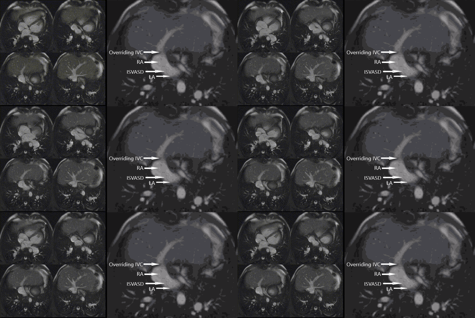
Movie 4: Cine- Axial stack showing IVC override of the atrial septum and ISVASD
Figure 4. Axial showing IVC override of the atrial septum and ISVASD.
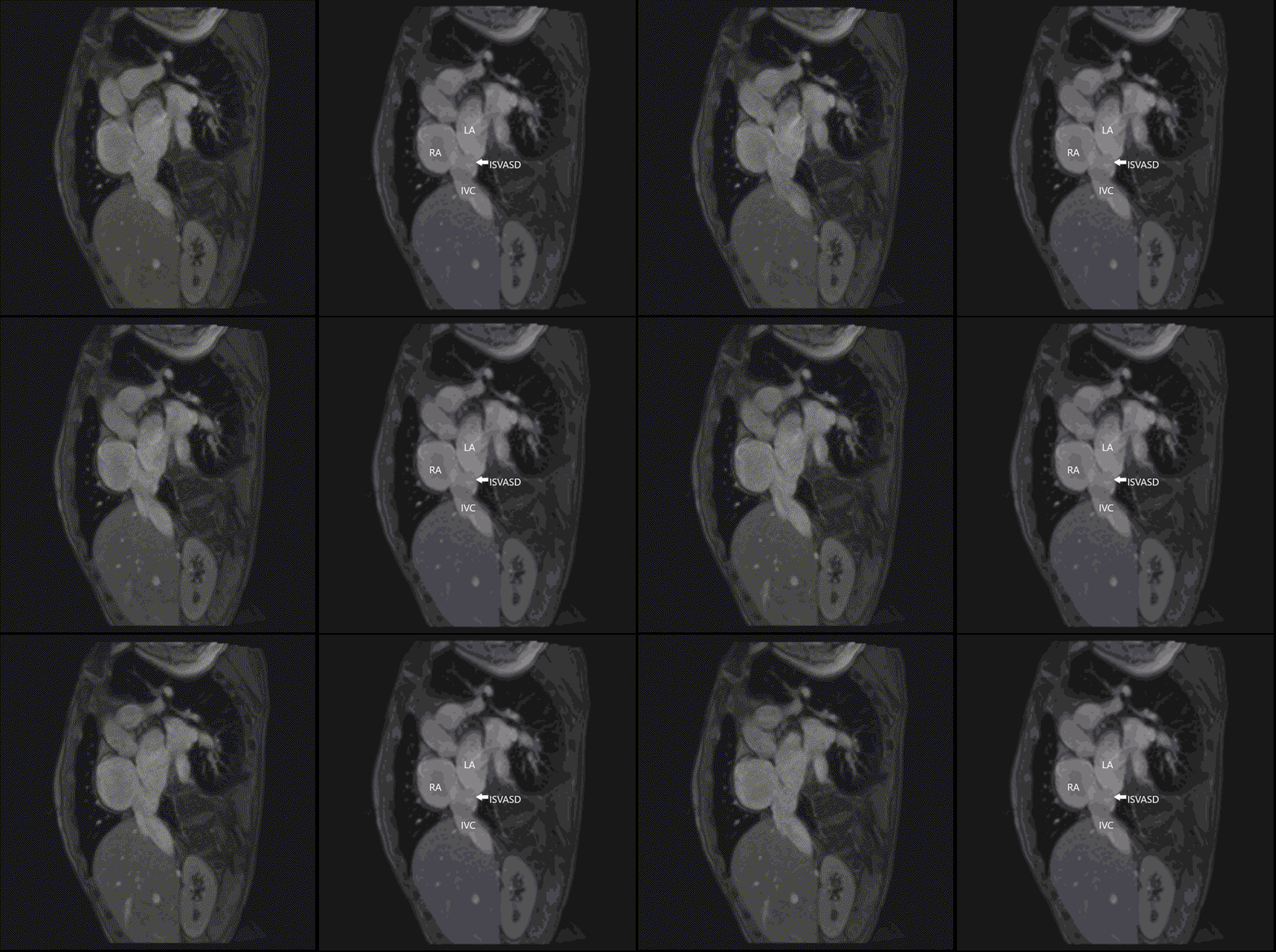
Figure 5. Bicaval view showing ISVASD

Figure 6. Phase contrast imaging with color overlay in the bicaval view showing the defect and left to right shunting at the defect
In addition, it showed dilated, tortuous, and engorged right pulmonary veins that formed a common confluence with a fold and drained into the left atrium (Movie 7, Figure 7).
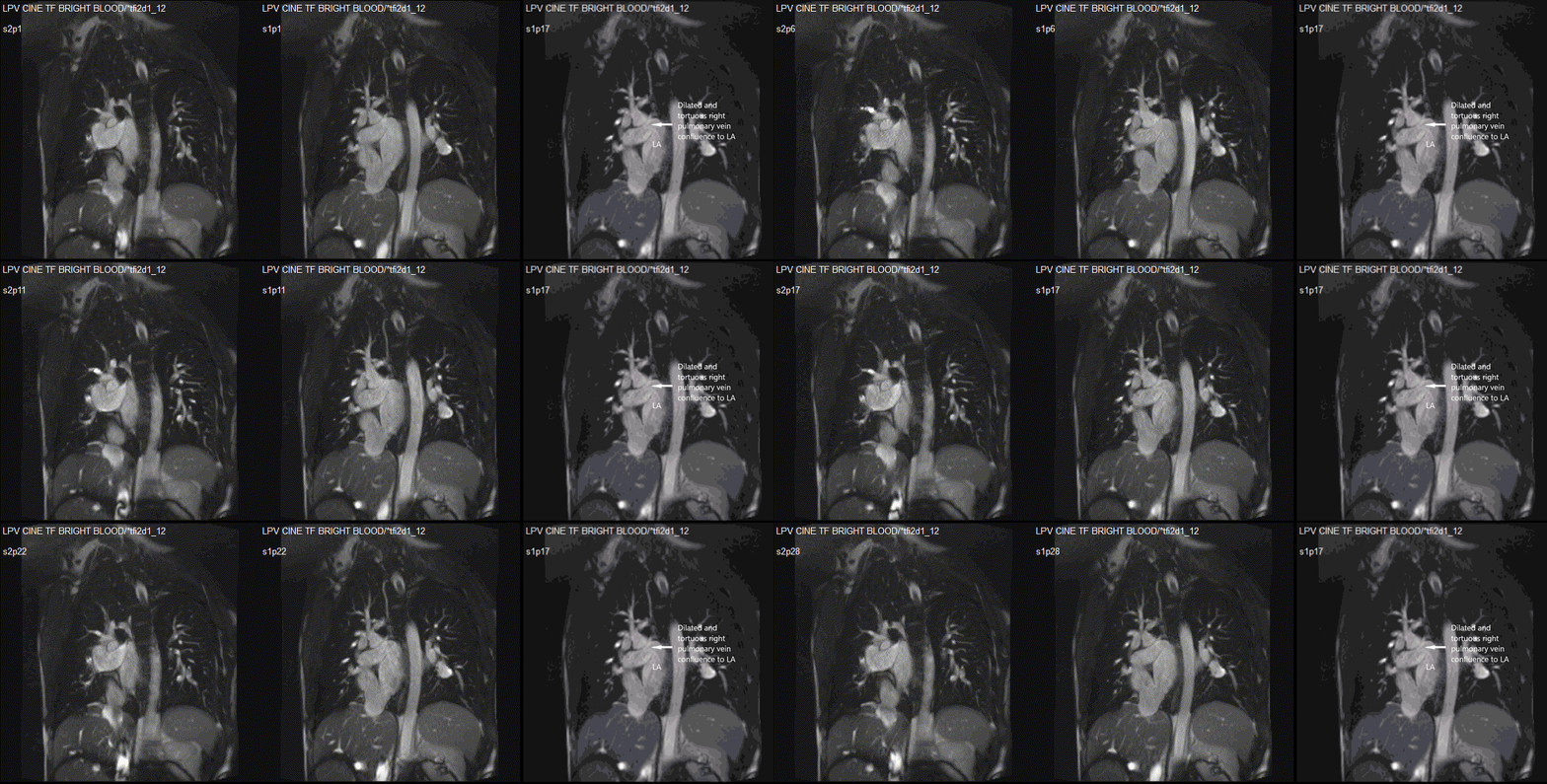
Figure 7. Dilated and tortuous right pulmonary vein confluence without obstruction
The left pulmonary veins formed a common pulmonary vein that drained to the left atrium, with flow turbulence of its junction suggestive of mild stenosis (Movie 8, Figure 8).
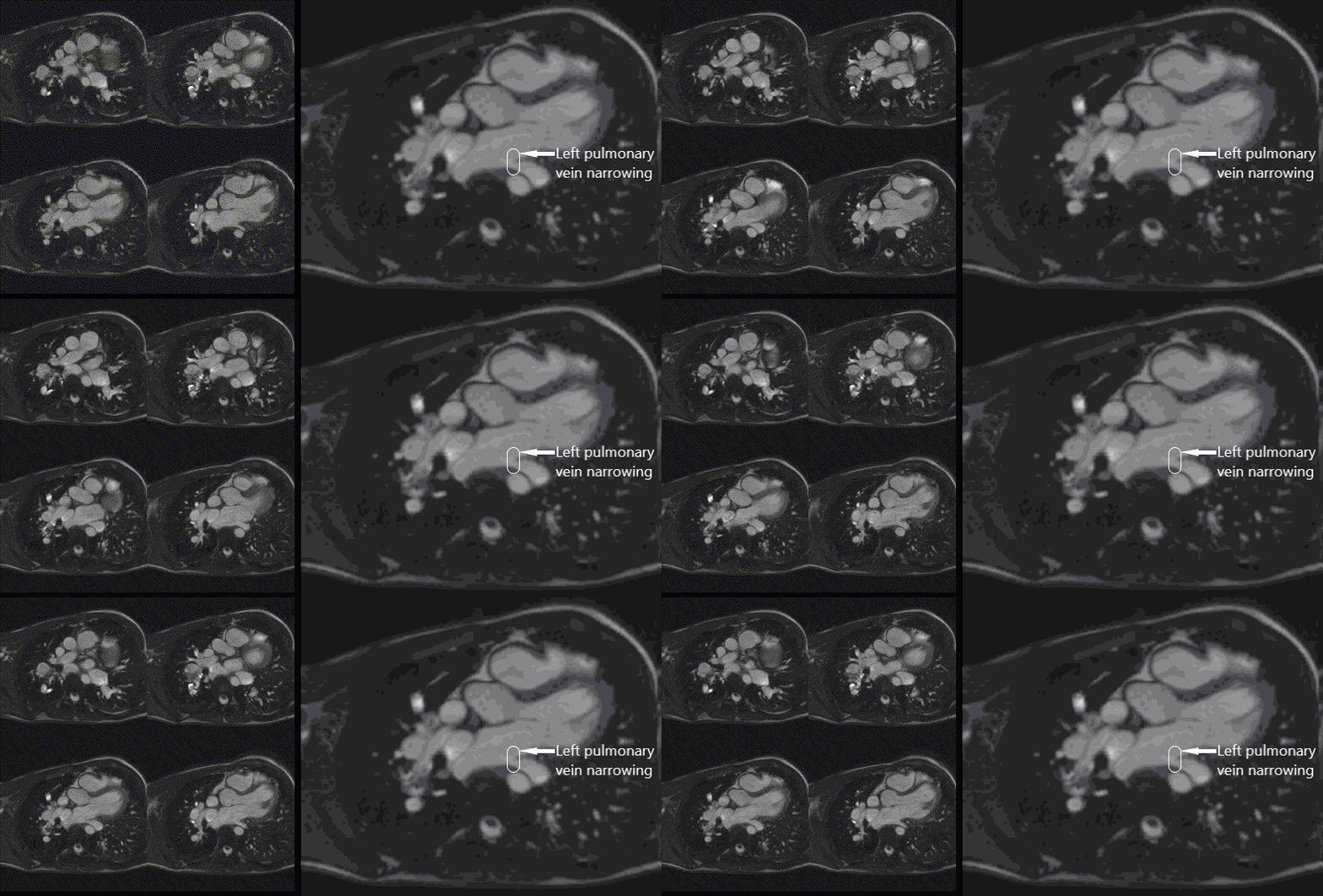
Figure 8. Slight narrowing of the left pulmonary vein entrance to the left atrium
The right atrium was mildly dilated with an area of 26 cm2, and the right ventricle was dilated with indexed RV volume of 150 ml/m2. The left atrium and ventricle were normal in size with an indexed left ventricular end diastolic volume of 85 ml/m2. There was preserved function with a left ventricular ejection fraction of 62% and a right ventricular ejection fraction of 64% (Movie 3). The Qp:Qs based on phase contrast imaging was 1.7. There was no left superior vena cava. Because of these findings and to evaluate pulmonary veins more thoroughly in anticipation of surgery, a cardiac catheterization was performed; it confirmed CMR findings of ISVASD with a Qp:Qs of 2:1. It is likely the shunt was higher by oximetry during catheterization due to supplemental oxygen use. There was no significant gradient across the anastomosis site both by wedge and direct pressure sampling from the veins. The left pulmonary veins appeared mildly narrow at the entrance into the left atrium. He had normal mean pulmonary artery pressure (14 mm Hg) and pulmonary vascular resistance (0.41 units). There was no restriction across the atrial septum. He underwent successful surgical repair with the intraoperative findings confirming the CMR findings.
Conclusion:
We report a rare case of ISVASD associated with TAPVR, which was missed at initial diagnosis and surgery. This resulted in continued right-sided dilation and atrial tachyarrhythmias. The association of TAPVR and ISVASD is extremely rare with this being the first case to the best of our knowledge based on literature search via PubMed. ISVASD is defined as a veno-atrial communication located posteriorly and inferiorly in the mouth of the inferior caval vein. (1–3) It accounts for about 3% of atrial septal defects (4) when looking at cardiac specimens, and 5% of sinus venosus defects. (5) ISVASD may also be associated to un-roofing of pulmonary veins into the IVC, and they can be associated to right lower lobe partial anomalous pulmonary venous return. (6)
References:
Case prepared by Associate Editor: Dr Sylvia Chen


