


Case from: M. Jay Campbell1,2, John Rhodes2, Stephen Darty1, Piers C.A. Barker1,2
Institute: 1Duke Cardiovascular Magnetic Resonance Center, Duke University Medical Center; 2Division of Pediatric Cardiology, Duke University School of Medicine
Clinical history: A previously healthy 23 year old military recruit presented to an outside hospital for a routine physical examination and a murmur was noted on exam. An echocardiogram with agitated saline injection was performed and contrast was immediately seen in the left atrium. The patient underwent cardiac catheterization and transesophageal echocardiogram which revealed concern for anomalous drainage of the right sided superior vena cava (SVC) to the left atrium. The patient was referred to our institution for further evaluation and management. On initial presentation, his systemic oxygen saturation was 90% on room air. The patient underwent cardiac MRI (CMR) and thoracic MRA to further evaluate his cardiac anatomy.
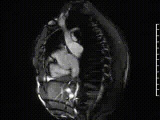
Movie 1

Movie 2
Description: CMR was performed on a 1.5T Siemens Avanto MRI scanner. SSFP images from a sagittal and coronal plane (Movie 1 and 2) reveal the presence of the right-sided SVC draining into the left atrium. There are two right-sided pulmonary veins draining into the right-sided SVC 7mm above the ostium of the right-sided SVC into the left atrium. A left-sided SVC is also seen.
Movie 3

Movie 4
Description: Movie 3 is a time resolved 3D angiogram with an injection of gadolinium contrast through an IV in the right arm. This reveals that the right-sided systemic venous drainage is through a right-sided SVC which empties into the left atrium. Movie 4 is a time resolved 3D angiogram with an injection of gadolinium contrast through an IV in the left arm. This reveals that the left-sided systemic venous drainage is through a left-sided SVC which empties into the coronary sinus and ultimately the right atrium.
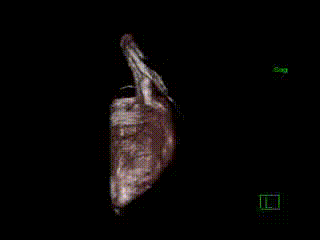
Movie 5
Description: Movie 5 is a 3D volume rendered reconstruction of an ECG gated contrast enhanced angiogram.
Velocity encoded imaging of the main pulmonary artery and the ascending aorta reveal a Qp/Qs of 0.75.
Conclusion:
CMR reveals the diagnosis of bilateral superior vena cavae with a left sided SVC draining into the coronary sinus and right atrium and an anomalous connection of the right sided SVC to the left atrium. The right upper and middle pulmonary veins attach to the right sided SVC and ultimately drain into the left atrium. There are no atrial septal defects seen. The connection of the right sided systemic venous drainage to the left atrium results in a right to left shunt.
Left sided SVC connecting to the coronary sinus is the most common systemic venous anomaly occurring in 0.3-0.5% of the general population.1,2 Right sided SVC connecting to the left atrium without an atrial septal defect is a rare anomaly with less than 30 previously reported cases. 2 This anomaly is usually associated with anomalous drainage of the right upper and middle pulmonary veins, as described in our case.3 Van Praagh et al. hypothesize that the embryological basis for this defect is deficiency of the common wall of the right sided SVC and right upper pulmonary vein.4 The result is unroofing of the right upper pulmonary veins into the right sided SVC. Preferential streaming of flow into the left atrium eventually results in involution on the proximal connection of the right sided SVC to the right atrium.4 3 cases of a right sided SVC draining to the left atrium with an associated left sided SVC have been described.3
An anomalous right sided SVC draining into the left atrium results in a right to left shunt. This can result in hypoxia, clubbing, risk of embolic events, and cerebral abscess. Our patient underwent successful surgical correction with ligation of the right sided SVC superior to the orifice of the anomalous pulmonary veins and reattachment of the right sided SVC to the right atrium with a Gore-Tex tube graft
Perspective:
This case illustrates a rare complex systemic venous anomaly and the use of cardiac MRI to define the anatomy. This defect can lead to life threatening complications and correct diagnosis is essential. The use of time resolved 3D angiography was instrumental in defining the anatomy in this complicated case. This case is an excellent illustration of the use of cardiac MRI in the diagnosis of complex congenital heart disease.
References:
- Perloff J. The clinical recognition of congenital heart disease. Philadelphia: W.B. Saunders Company, 1994; p703.
- Hong S, Nayar A, Srichai M, Morgan J, Meyer D, Katz E. A case of anomalous superior vena cava with anomalous pulmonary veins-when two wrongs do not make a right. Echocardiography 2011; 28: E39-E41.
- Van Praagh S, Geva T. Abnormal systemic venous connections. In: Allen H, Driscoll D, Shaddy R, Feltes T. Moss and Adams’: Heart disease in infants, children, and adolescents 2008. 792-817
- Van Praagh S, Geva T, Lock J.E., del Nido P.J., Vance M.S., Van Praagh R. Biatrial or left atrial drainage of the right superior vena cava: anatomic, morphogenetic and surgical considerations. Pediatric Cardiology 2003; 24: 350-363.
- Sadek H, Gilkeson R, Hoit B, Brozovich F. Case of anomalous right superior vena cava. Circulation 2006; 114:e532-e533.
- Sajja LR, Koneti NR, Mannam G, Sundaram MK. Correction of anomalous drainage of right superior vena cava to left atrium. Asian Cardiovasc Thorac Ann. 2008;16(4):e32-4.
COTW handling editor: Micheal Jay Campbell