


Case from: Sebastiaan Hammer, MD, PhD1; A. A. W Roest, MD, PhD2; Albert de Roos, MD, PhD1
Institute: 1 Department of Radiology, Leiden University Medical Centre, Leiden, The Netherlands; 2 Department of Pediatric Cardiology, Leiden University Medical Centre, Leiden, The Netherlands
Clinical history:
A 24 year old man underwent surgical repair for tetralogy of Fallot (TOF) with a pulmonary valvotomy at the age of 10 months. He presented in 2006 with increased complaints of shortness of breath during exercise. An EKG was obtained, showing QRS prolongation (178 ms), due to chronic right ventricular overload and a right-bundle branch block (after closure of the VSD, Figure 1). Cardiovascular magnetic resonance (CMR) was performed to assess right ventricular function, as well as flow velocities across the pulmonary valve.

Figure 1
CMR Findings:
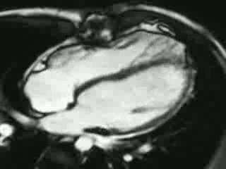
CMR showed the classic findings of grown up congenital heart disease (GUCHD) in patients with corrected TOF. As the stenotic pulmonary valve is surgically dilated, over the years progressive pulmonary insufficiency develops frequently. CMR showed a dilated (box-shaped) right ventricle, with paradoxal septal diastolic movement in the 4-chamber cine view (Figure 2 and Movie 1). It also suggests mild right ventricular hypertrophy.


Figure 2 & Movie 1
A regurgitant jet is visualized at the level of the pulmonary valve (velocity mapping; Movie 2). The corresponding flow versus time curve is shown in Figure 3 (highlighted area depicts backward flow of 85 ml, forward flow was 175 ml). A pulmonary insufficiency of 49% was documented. In addition, note the end-diastolic forward flow, indicating right ventricular restrictive physiology. Right ventricular systolic function was quantified using dedicated software (Mass), by drawing endocardial contours on end-diastolic and end-systolic transversal cine images. End diastolic volume (EDV) was 447 ml or 235ml/m2, end systolic volume (ESV) 266 ml or 140ml/m2, resulting in an ejection fraction of 40%. After correction for pulmonary insufficiency, the effective ejection fraction was 21% (quantified as (EDV – regurgitation flow) – ESV/ EDV).


Movie 2 & Figure 3
The patient ultimately was re-operated in 2007 and a pulmonary homograft was placed. Figure 4 is a flow vs. time curve obtained 3 years later which reveals no pulmonary insufficiency. Figure 5 and Movie 3 show the improvements in right ventricular dimensions 3 years later. Movie 1 is shown again for direct comparison. The patient’s exercise capacity improved concomitantly. EDV decreased to 263 ml or 138ml/m2, ESV was 159 ml or 84ml/m2; in the absence of pulmonary insufficiency, the effective ejection fraction increased to 40%. Table 1 illustrates right ventricular volumes and ejection fraction pre- and post-operatively.


Figure 4 & Figure 5
Movie 1: Pre-operative & Movie 3
Conclusion:
The present case shows the classic findings of GUCHD in surgically corrected patients with TOF. Pulmonary insufficiency is a well-known disease entity in operated patients with TOF. As a consequence, right ventricular pressure rises, which results in increased end-diastolic volumes, i.e. dilatation of the right ventricle. Pulmonary valve replacement improves right ventricular dimensions and patient complaints, as can be objectified by improved New York Heart Association Classification (NYHA). This patient’s NYHA class improved from class 2 pre-operatively to class 1 post-operatively. Using CMR, adequate estimates of RV dimensions, ejection fraction and pulmonary insufficiency can be obtained.
Perspective:
CMR can play an important role in determining right ventricular overflow and monitor improvements after surgical repair in patients with GUCHD. Normalization of right ventricular dimensions have been documented when pre-operative, CMR documented, RV end-diastolic volumes are below 160 mL/m2.
References:
1. Gatzoulis et al. Mechanoelectrical interaction in tetralogy of Fallot. Circulation. 1995 Jul 15; 92(2):231-7.
2. Gaca et al. Repair of congenital heart disease, a primer. Part 2. Radiology. 2008 Jul; 248(1). 44-60.
3. Oosterhof et al. Preoperative thresholds for pulmonary valve replacement in patients with corrected tetralogy of Fallot using cardiovascular magnetic resonance. Circulation. 2007 Jul; 116(5). 545-51.
4. Henkens et al. Predicting outcome of pulmonary valve replacement in adult tetralogy of Fallot patients. Ann Thoracic Surg. 2007 Mar; 83(3). 907-11.
5. Kalra N, Klewer SE, Raasch H, Sorrell VL. Update of tetralogy of Fallot for the adult cardiologist including a brief historical and surgical perspective. Congent Heart Dis. 2010 May-June; 5(3):208-19.
Submit your case here
COTW handling editor: M. Jay Campbell, MD
Have your say: What do you think? Latest posts on this topic from the forum