Case from: Amr M. Ajlan, Jonathan Leipsic, Cameron Hague
Institute: Cardiac Imaging Unit, St. Paul’s Hospital, Vancouver, BC, Canada
Clinical history: 46-year-old male patient referred from the echocardiography laboratory to evaluate a known atrial septal defect (ASD) and to assess the status of the mitral valve. CMR was used to accurately evalute the degree of the shunt and the size of the RV.
CMR Findings:
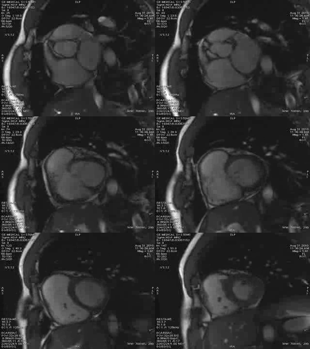
Movie 1
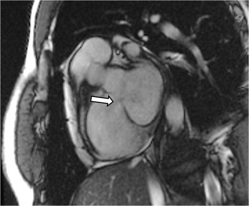
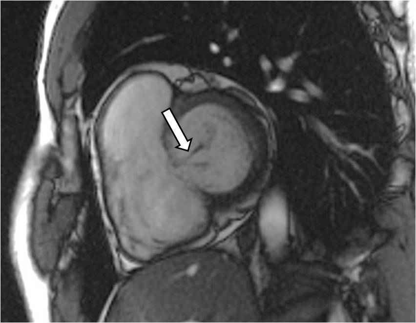
Figure 1 & Figure 2
Left ventricular short axis SSFP cine: Movie.1 shows a low interatrial communication, in keeping with a primum-type ASD. A left-to-right dephasing jet is seen during atrial contraction. A ventricular systolic posteriorly directed mitral dephasing jet is also seen, in keeping with mitral regurgitation. Still frame images are selected to delineate the ASD-related left-to-right (arrow in Figure.1) and mitral regurgitation (arrow in Figure.2) jets
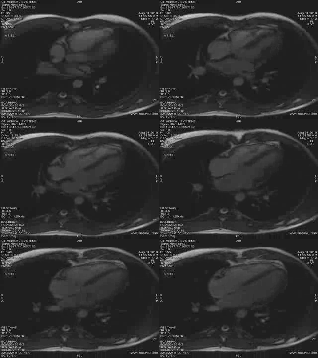
Movie 2
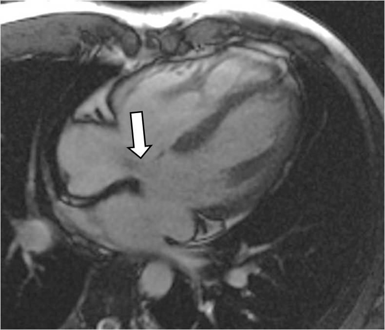
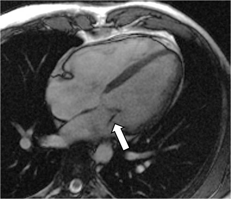
Figure 3 & Figure 4
Four-chamber SSFP cine shows the primum ASD, but also demonstrates the mitral leaflets motion and their overall morphological appearance. This sequence clearly shows a sizable anterior mitral leaflet cleft, which opposes during valve closure. Still frame images are selected to delineate the ASD-related left-to-right jet (arrow in Figure.3) and the anterior mitral leaflet cleft (arrow in Figure.4).
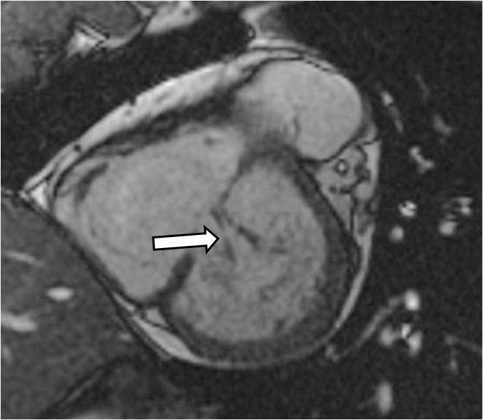
Figure 5
Still frame image from thin slice SSFP acquisition in the plane of the mitral valve (Figure.5): This image further delineates the cleft of the anterior mitral valve (arrow in Figure.5).
Conclusion: The CMR confirmed the presence of a primum ASD and showed an associated anterior mitral leaflet cleft. Calculations using velocity-encoded phase contrast images over the aorta and pulmonary artery (not shown) revealed a left-to-right shunt ratio (Qp/Qs) of 1.7. The degree of mitral regurgitation was mild to moderate.
Perspective: When facing a case of primum ASD, meticulous search for an associated anterior mitral leaflet cleft should be performed. Transthoracic or transesophageal Echo are the classic imaging methods of choice. This case highlights that CMR may be an additional excellent modality for such an assessment. Adding thin slice (4-6 mm) SSFP cine images along the mitral valve plane can increase the ability to characterise the leaflets morphology. The presence of a mitral leaflet cleft has important implications when ASD surgical correction is attempted and should be addressed accordingly. This is because the mitral valve leaflet cleft usually causes significant mitral valve regurgitation. The size of the RV and LV should be measured by CMR before and after surgery. The left-to-right shunt usually causes an enlarged RV, the mitral regurgitation an enlarged LV. Both ventricles are usually expected to become smaller after successful surgery.
It is important to note that some prefer the term “left-sided atrioventricular valve” over the term “mitral valve” in cases of a primum ASD. This is because a primum ASD is a atrioventricular septal defect that is accompanied by various forms of AV valve deformation leading to AV valves with up to seven leaflets. Accordingly, some name the “mitral valve cleft” an “additional commissure of the left-sided atrioventricular valve”. Therefore, however far more seldom, a primum ASD may be accompanied by “tricuspid valve cleft” or an “additional commissure of the right-sided atrioventricular valve”.
References:
Gutiérrez FR, Ho ML, Siegel MJ. Practical applications of magnetic resonance in congenital heart disease. Magn Reson Imaging Clin N Am. 2008 Aug;16(3):403-35.
Tamura M, Menahem S, Brizard C. Clinical features and management of isolated cleft mitral valve in childhood. J Am Coll Cardiol. 2000 Mar 1;35(3):764-70.
Peter B. Manning. Operative Techniques in Thoracic and Cardiovascular Surgery: A Comparative Atlas. Autumn 2004 (Vol. 9, Issue 3, Pages 240-246).
Yuan SM, Shinfeld A, Raanani E. Mitral valve cleft associated with secundum atrial septal defect: case report and review of the literature. Monaldi Arch Chest Dis. 2007 Mar;68(1):48-51.
Jemielity M, Perek B, Paluszkiewicz L, Dyszkiewicz W. Results of surgical repair of ostium primum atrial septal defect in adult patients. J Heart Valve Dis. 2001 Jul;10(4):525-9.
Submit your case here
COTW handling editor: Sohrab Fratz
Have your say: What do you think? Latest posts on this topic from the forum


