SCMR

Mystery revealed: It takes Cardiovascular Magnetic Resonance to diagnose Inferior Sinus Venosus Defect in an adult status post Total Anomalous Pulmonary Venous Return repair

González Estévez N1 , Priya S2, Ashwath ML 2, Ashwath RC 1

1Stead Family Department of Pediatrics, 2University of Iowa Hospitals and Clinics, University of Iowa Health Care

Clinical history:

The patient is a 27-year-old male status post complete surgical repair of supra-cardiac total anomalous pulmonary venous return (TAPVR) in infancy. He was followed intermittently until 15 years old, with no concerning imaging findings, and was essentially asymptomatic. At 23 and 27 years of age, he presented to the emergency department with atrial fibrillation and atrial flutter, respectively, requiring cardioversion. He had a transthoracic echocardiogram (TTE) performed which showed concerning findings of a mildly dilated inferior vena cava (IVC) and hepatic veins with exaggerated reversal of flow (Movie 1, Figure 1), dilated right ventricle (RV), abnormal venous flow pattern in the right atrium (Movie 1), and a dilated venous confluence (Movie 2, Figure 2)
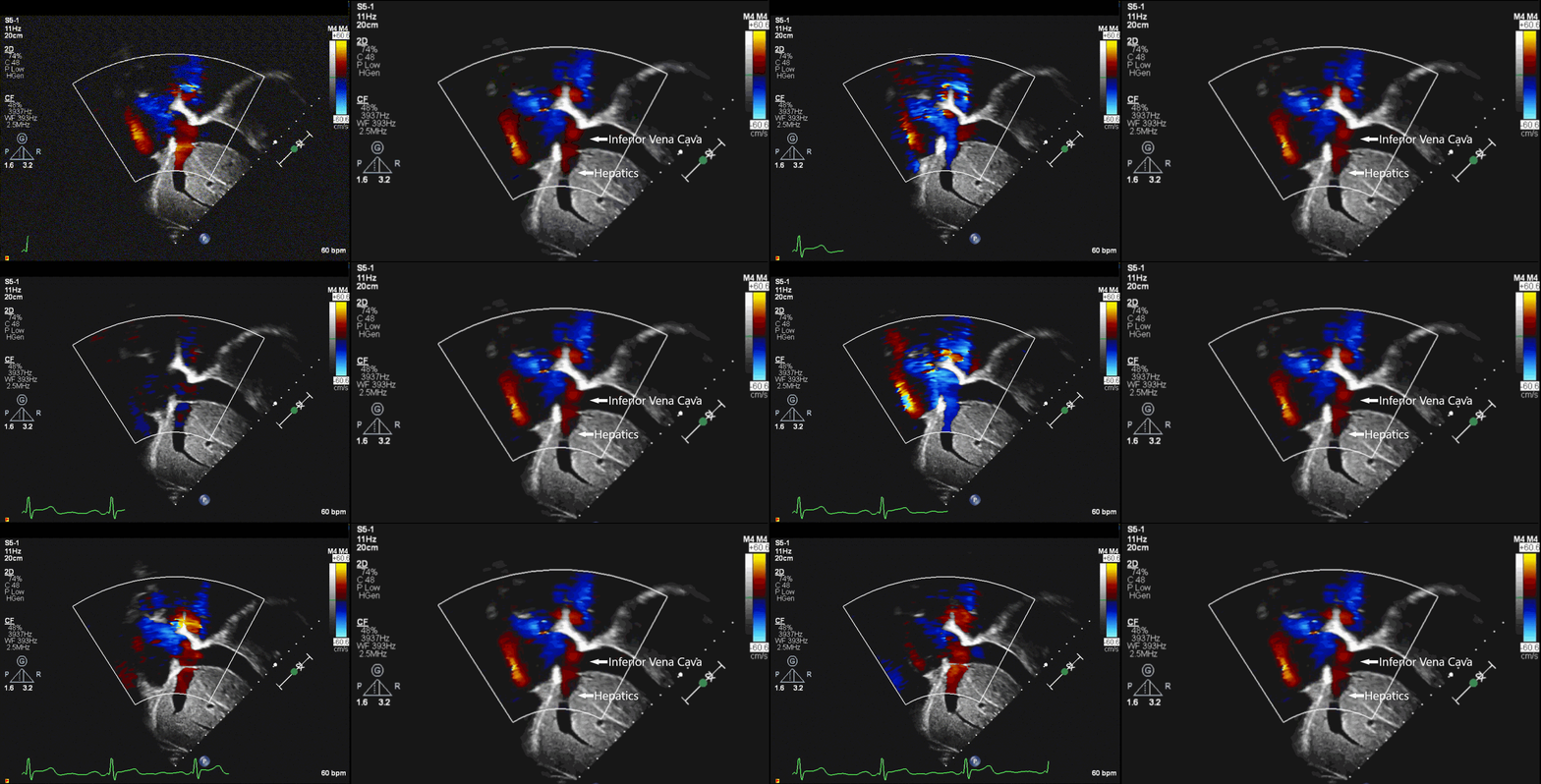
Movie 1: Bicaval view showing a dilated IVC and hepatics with flow reversal. Turbulent venous flow into the right atrium is also evident.

Figure 1. Bicaval view showing a dilated IVC and hepatics with flow reversal. 
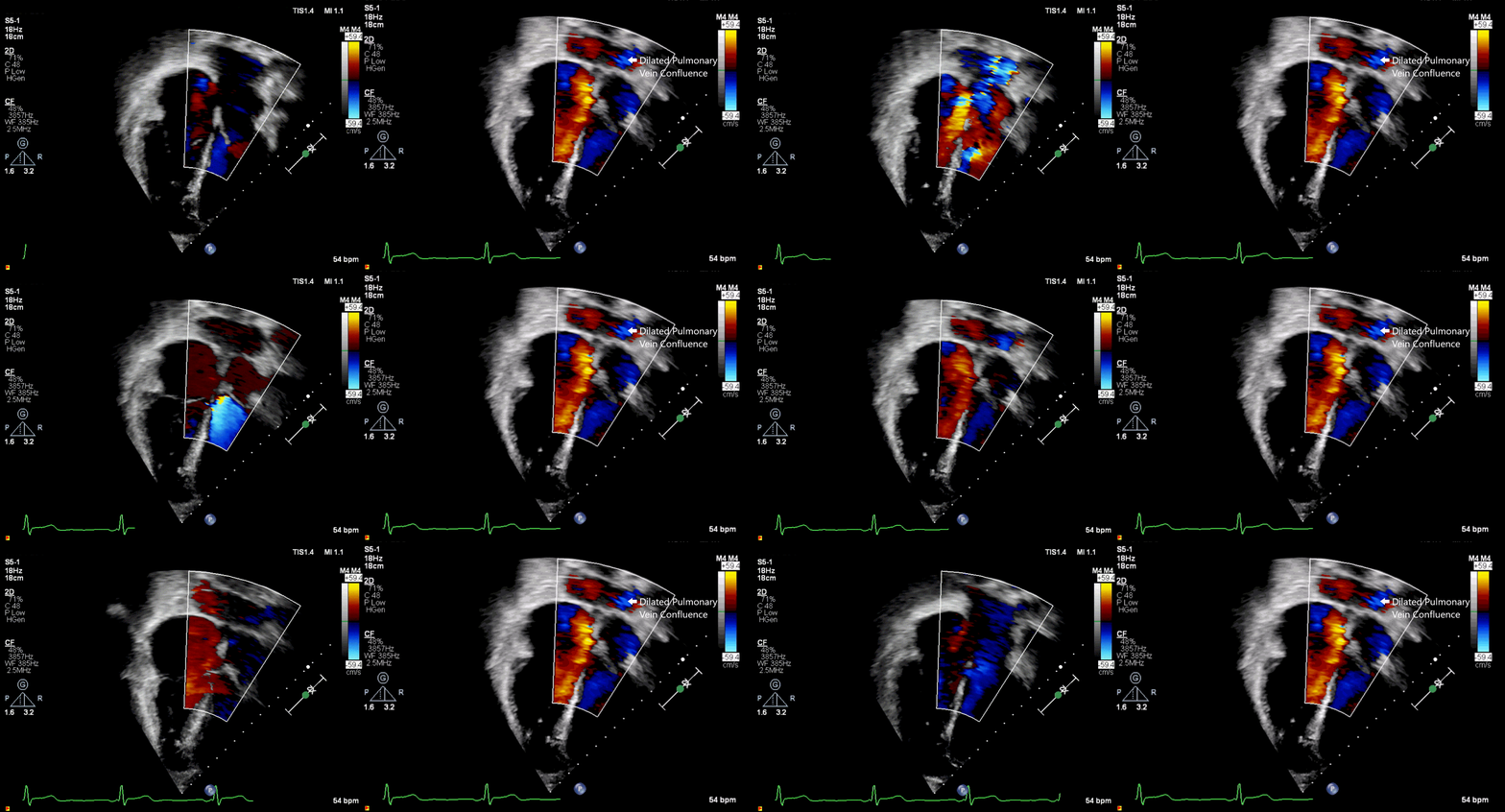
Movie 2: Color flow mapping in apical four chamber view showing the dilated pulmonary vein confluence.

Figure 2. Color flow mapping in apical four chamber view showing the dilated pulmonary vein confluence.

CMR Findings:

A cardiac magnetic resonance imaging (CMR) study was performed to assess pulmonary veins, venous confluence, and unexplained right ventricular dilation (Movie 3, Figure 3). 
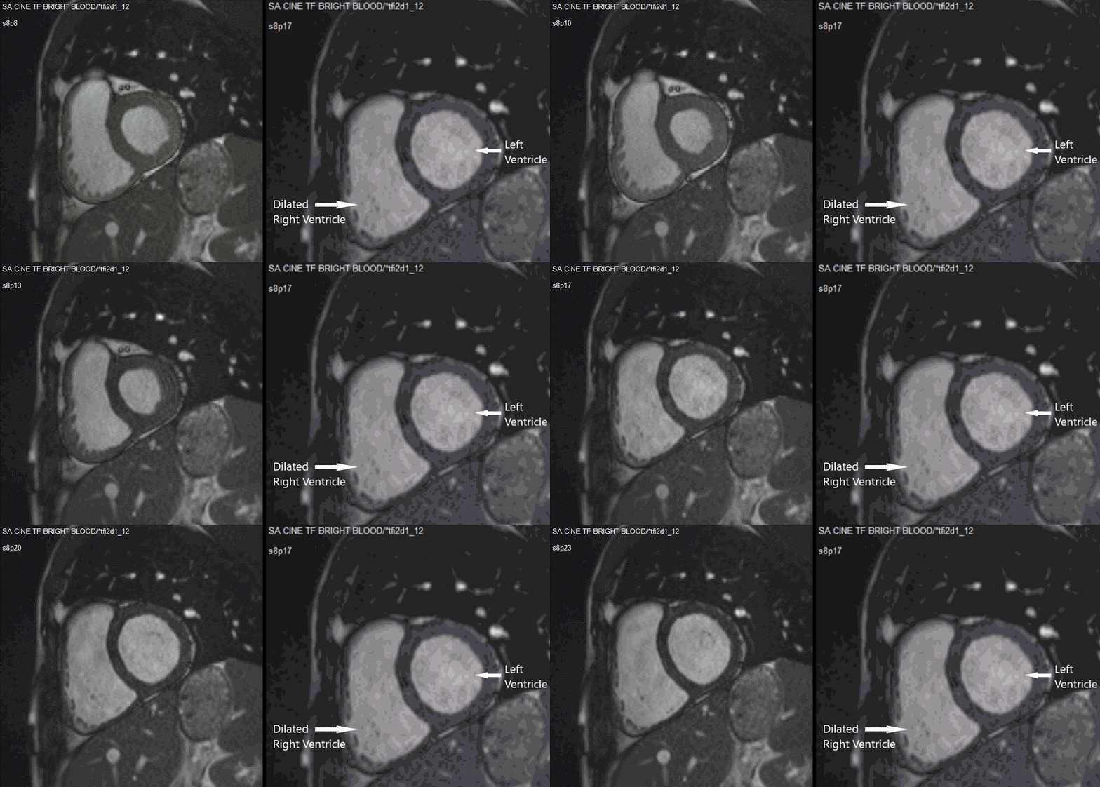
Movie 3: Cine- Short axis showing severe right ventricular dilation 
Figure 3. Short axis showing severe right ventricular dilation

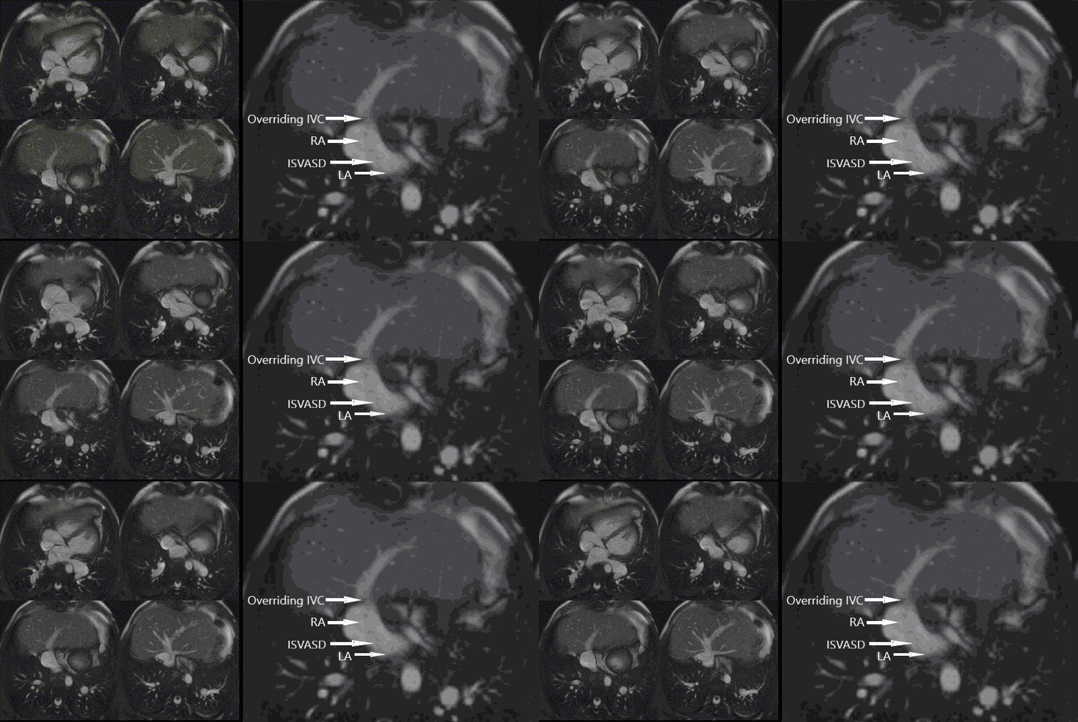
Movie 4: Cine- Axial stack showing IVC override of the atrial septum and ISVASD

Figure 4. Axial showing IVC override of the atrial septum and ISVASD.  
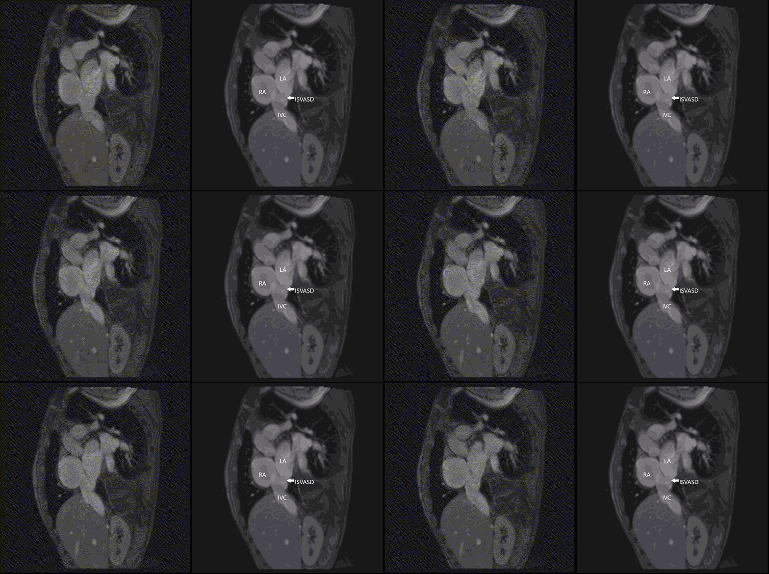
Movie 5: Cine- Bicaval view showing ISVASD

Figure 5. Bicaval view showing ISVASD
Movie 6: Phase contrast imaging with color overlay in the bicaval view showing the defect and left to right shunting at the defect


Figure 6. Phase contrast imaging with color overlay in the bicaval view showing the defect and left to right shunting at the defect 
 

In addition, it showed dilated, tortuous, and engorged right pulmonary veins that formed a common confluence with a fold and drained into the left atrium (Movie 7, Figure 7). 
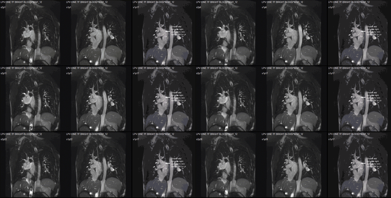
Movie 7: Cine- Dilated and tortuous right pulmonary vein confluence without obstruction


Figure 7.
 Dilated and tortuous right pulmonary vein confluence without obstruction  

The left pulmonary veins formed a common pulmonary vein that drained to the left atrium, with flow turbulence of its junction suggestive of mild stenosis (Movie 8, Figure 8). 
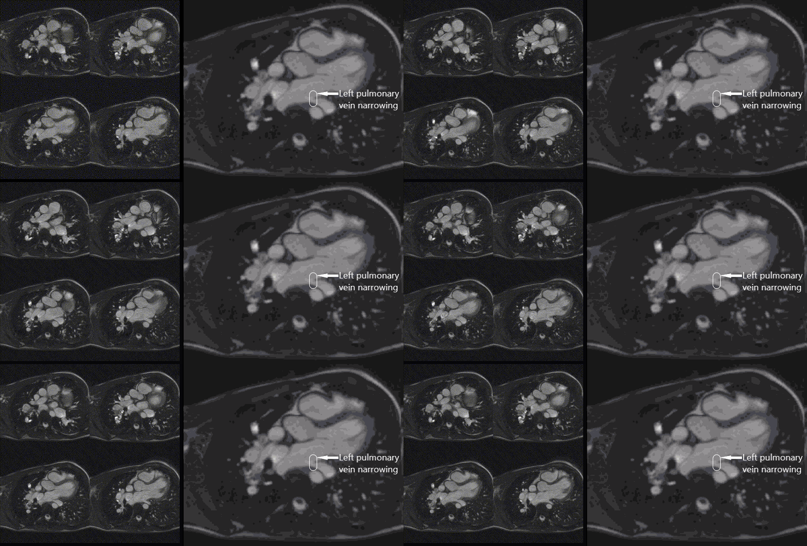
Movie 8: Cine- Slight narrowing of the left pulmonary vein entrance to the left atrium


Figure 8.
 Slight narrowing of the left pulmonary vein entrance to the left atrium  

The right atrium was mildly dilated with an area of 26 cm2, and the right ventricle was dilated with indexed RV volume of 150 ml/m2. The left atrium and ventricle were normal in size with an indexed left ventricular end diastolic volume of 85 ml/m2. There was preserved function with a left ventricular ejection fraction of 62% and a right ventricular ejection fraction of 64% (Movie 3). The Qp:Qs based on phase contrast imaging was 1.7. There was no left superior vena cava. Because of these findings and to evaluate pulmonary veins more thoroughly in anticipation of surgery, a cardiac catheterization was performed; it confirmed CMR findings of ISVASD with a Qp:Qs of 2:1. It is likely the shunt was higher by oximetry during catheterization due to supplemental oxygen use. There was no significant gradient across the anastomosis site both by wedge and direct pressure sampling from the veins. The left pulmonary veins appeared mildly narrow at the entrance into the left atrium. He had normal mean pulmonary artery pressure (14 mm Hg) and pulmonary vascular resistance (0.41 units). There was no restriction across the atrial septum. He underwent successful surgical repair with the intraoperative findings confirming the CMR findings. 

Conclusion: 

CMR is an excellent non-invasive imaging modality to accurately diagnose ISVASD and to clearly delineate pulmonary venous anatomy, including any obstruction. It also helps to calculate the shunt fraction. We feel that CMR is highly beneficial in patients with an uncertain diagnosis and a high index of suspicion with abnormal findings in TTE for accurate preoperative diagnosis. 
 
Perspective: 

We report a rare case of ISVASD associated with TAPVR, which was missed at initial diagnosis and surgery. This resulted in continued right-sided dilation and atrial tachyarrhythmias. The association of TAPVR and ISVASD is extremely rare with this being the first case to the best of our knowledge based on literature search via PubMed.  ISVASD is defined as a veno-atrial communication located posteriorly and inferiorly in the mouth of the inferior caval vein. (1–3) It accounts for about 3% of atrial septal defects (4) when looking at cardiac specimens, and 5% of sinus venosus defects. (5) ISVASD may also be associated to un-roofing of pulmonary veins into the IVC, and they can be associated to right lower lobe partial anomalous pulmonary venous return. (6)  

 
ISVASD are difficult to diagnose. Banka et al. compared surgical outcomes of ISVASD with those of large secundum ASD and found that 64% of patients with ISVASD were misdiagnosed by preoperative imaging and 40% were misdiagnosed on operative assessment. The authors did note that preoperative diagnostic accuracy has continued to improve over time. They also found that the ISVASD group had worse technical outcomes, a higher rate of reinterventions, longer durations of cardiopulmonary bypass, Intensive Care Unit stay, and hospitalization than the secundum ASD group. (7) Their results correlated with a prior smaller series by Crystal et al. that found incomplete preoperative diagnosis in 55% of their patients with ISVASD. However, this earlier series reported excellent outcomes with no re-intervention or residual defects reported. (3) Based on these studies, adequate diagnosis and delineation of these defects is crucial for proper surgical planning.  
 
Diagnosis of ISVASD historically has relied on transthoracic and transesophageal echocardiography (TEE) as well as cardiac catheterization. These techniques come with their inherent disadvantages: for TEE, these include limited acoustic windows in adults and certain body types, need for sedation, and limited availability; for cardiac catheterization disadvantages include radiation exposure, need for sedation, and the invasive nature of the procedure. More recently, however, the use of CMR for evaluation of these defects has become more common. (8) Valente et al. have proposed the use of CMR as a non-invasive alternative to previously used evaluations, as they found that surgical repair confirmed CMR accuracy in diagnosis of sinus venosus defects and even identified unsuspected additional abnormalities while providing other crucial anatomic and hemodynamic information for surgical planning. (8)   
Our patient presented after an arrhythmia, which led to further evaluation with TTE. Non-diagnostic, yet concerning findings of a dilated IVC and hepatics with flow reversal—as well as an enlarged pulmonary vein confluence and unexplained right ventricular dilation—prompted escalation to CMR, and diagnosis was confirmed. CMR was crucial in diagnosis of this inferior sinus venosus atrial septal defect that had gone undiagnosed for 27 years despite extensive cardiac evaluation, surgery, and follow up for total anomalous pulmonary venous return. CMR remains the gold standard in the evaluation of such rare defects and carries the added advantage of obtaining good resolution of extracardiac structures and pulmonary venous anatomy, as well as flow data shunt fractions. This allowed for accurate diagnosis and thus facilitated appropriate further surgical management for our patient.
 

References:

1. Ettedgui JA, Siewers RD, Anderson RH, Park SC, Pahl E, Zuberbuhler JR. Diagnostic echocardiographic features of the sinus venosus defect. Br Heart J. 1990 Nov;64(5):329–31.

2.  Attenhofer Jost CH, Connolly HM, Danielson GK, Bailey KR, Schaff HV, Shen W-K, et al. Sinus venosus atrial septal defect: long-term postoperative outcome for 115 patients. Circulation. 2005 Sep 27;112(13):1953–8.

3. Crystal MA, Al Najashi K, Williams WG, Redington AN, Anderson RH. Inferior sinus venosus defect: echocardiographic diagnosis and surgical approach. J Thorac Cardiovasc Surg. 2009 Jun;137(6):1349–55.

4. Muñóz-Castellanos L, Espinola-Zavaleta N, Kuri-Nivón M, Ruíz JF, Keirns C. Atrial septal defect: anatomoechocardiographic correlation. J Am Soc Echocardiogr Off Publ Am Soc Echocardiogr. 2006 Sep;19(9):1182–9.

5.  Hidalgo A, Ho M-L, Bhalla S, Woodard PK, Billadello JJ, Gutierrez FR. Inferior type sinus venosus atrial septal defect: MR findings. J Thorac Imaging. 2008 Nov;23(4):266–8.

6.  Snarr BS, Liu MY, Zuckerberg JC, Falkensammer CB, Nadaraj S, Burstein D, et al. The Parasternal Short-Axis View Improves Diagnostic Accuracy for Inferior Sinus Venosus Type of Atrial Septal Defects by Transthoracic Echocardiography. J Am Soc Echocardiogr Off Publ Am Soc Echocardiogr. 2017 Mar;30(3):209–15.

7.  Banka P, Bacha E, Powell AJ, Benavidez OJ, Geva T. Outcomes of inferior sinus venosus defect repair. J Thorac Cardiovasc Surg. 2011 Sep;142(3):517–22.

8.  Valente AM, Sena L, Powell AJ, Del Nido PJ, Geva T. Cardiac magnetic resonance imaging evaluation of sinus venosus defects: comparison to surgical findings. Pediatr Cardiol. 2007 Feb;28(1):51–6.

 Case prepared by Associate Editor: Dr Sylvia Chen

Newsbeat Banner

SCMR Newsletter brought to you by NewsBeat

Read the current SCMR NewsBeat to stay up to date with the latest SCMR News!