






Sam Younes, MD, Daniel Davies, MD, Francis Ezekwueme, MD, Anita Radhakrishnan, MD
Cardiovascular Institute, Allegheny General Hospital, Pittsburgh, PA, USA
Clinical History
A 75-year-old male had sudden onset chest pain and left shoulder numbness on exertion which prompted the patient to call for emergency medical care. Initial electrocardiogram (ECG) showed ST elevations in leads II, III, AVF, V5 and V6 and ST depressions in V1-V3. On arrival at a primary percutaneous coronary intervention center, repeat ECG demonstrated findings consistent with inferolateral myocardial infarction. High sensitivity troponin was elevated at 3500 ng/L (normal range: 0-22 ng/L). He first had a computed tomography angiogram (CTA) of the chest, abdomen and pelvis completed to rule out dissection given the finding of widened mediastinum on chest x-ray at which time a left ventricular (LV) aneurysm was seen (Figure 1). He then underwent left heart catheterization (LHC) with drug eluting stent placement in an obtuse marginal branch. The patient had a repeat LHC the next day and had another drug eluting stent placed in an 80% occluded mid-left anterior descending artery and sub-total occluded right coronary artery. He then underwent cardiovascular magnetic resonance (CMR) imaging for evaluation of the aneurysm and myocardial viability. The patient was discharged home a few days later and followed up with outpatient cardiology. One year later, the patient presented to undergo repeat CMR to evaluate the LV aneurysm.
 |
| Figure 1. CTA image of the LV aneurysm at initial presentation. |
CMR Findings
CMR studies were performed both at initial presentation and at one year follow up, both using a 1.5 Tesla Magnetom Aera system (Siemens Healthineers, Erlangen Germany). A series of cine balanced steady state free precession (bSSFP) imaging performed in 2, 3, 4-chamber projections, T1 (black blood) morphology, triple inversion recovery T2 imaging, LGE imaging acquired in 2, 3, 4-chamber and short axis planes, first pass perfusion imaging acquired in 2, 3, 4-chamber and short axis planes and phase contrast imaging were performed for both the initial and repeat CMR scans. The follow-up CMR also had long inversion time (TI) sequence imaging.
The first CMR scan revealed a reduced left ventricular ejection fraction (LVEF) of 33% (normal range: 57-77%) with a LV end-diastolic volume (LVEDV) of 187 mL (normal range: 106-214 mL), LV end-diastolic volume index (LVEDVI) of 103 mL/m2 (normal range: 57-105 mL/m2), LV end-systolic volume (LVESV) of 121 mL (normal range: 26-82 mL) and LV end-systolic volume index (LVESVI) of 69 mL/m2 (normal range: 14-38 mL/m2). He had a right ventricular ejection fraction (RVEF) of 63% (normal range: 52-72%) with a right ventricular (RV) end-diastolic volume (RVEDV) of 85 mL (normal range: 118-250 mL), RV end-diastolic volume index (RVEDVI) of 48 mL/m2 (normal range: 61-121 mL/m2), RV end-systolic volume (RVESV) of 32 mL (normal range: 41-117 mL) and a RV end-systolic volume index (RVESVI) of 18 mL/m2 (normal range: 19-59 mL/m2).
The initial CMR scan revealed a 35 x 35 mm basal lateral myocardial aneurysm which correlated with the CTA (Figures 1 and 2). The tissue involving the wall of the aneurysm had LGE with microvascular obstruction consistent with inferolateral infarcted myocardium (Figures 3 and 4). First pass perfusion imaging was performed to assess myocardial blood delivery and extent of flow limiting disease. After revascularization, the presence of microvascular obstruction represents an unfavorable prognosis for salvageable myocardium. T1 and T2 mapping was not performed.
The second CMR scan revealed a persistently reduced LVEF of 33% with LVEDV of 210 mL, LVEDVI of 109 mL/m2, LVESV of 140 mL and LVESVI of 73 mL/m2. RVEF remained normal at 69% with a RVEDV of 100 mL, RVEDVI of 52 mL/m2, RVESV of 31 mL and RVESVI of 16 mL/m2. The scan also revealed an increase in size of the LV aneurysm to 49 x 42 mm with a new irregular outpouching along the basal anterolateral wall, measuring 15 x 10 mm (Figures 5 and 6, Movies 1 and 2). There was a similar LGE pattern as in the initial scan (Figure 6). This is representative of persistent transmural scarring. Long TI sequence imaging was also performed and was negative for LV mural thrombus. First pass perfusion was performed and revealed a resting perfusion defect of the apex and basal inferolateral wall, consistent with the infarcted territory. T1 and T2 mapping was also not performed on the second CMR scan.
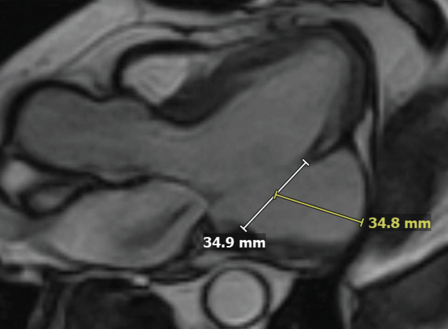 |
| Figure 2. Three chamber bSSFP image at peak systole of the LV aneurysm at initial presentation. This cross section allowed for the best direct visualization of the aneurysm for tissue characterization and measurement. |
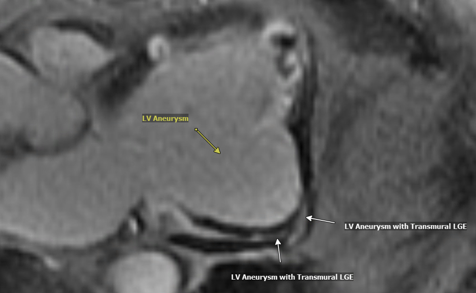 |
| Figure 3. Three chamber phase sensitive inversion recovery (PSIR) image of the LV aneurysm seen at initial presentation. The LGE pattern is representative of microvascular obstruction and transmural LGE in the area of the aneurysm. |
 |
| Figure 4. Short axis basal PSIR of the LV aneurysm on initial presentation showing transmural LGE. |
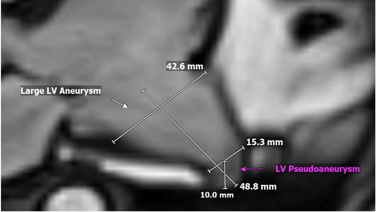 |
| Figure 5. Three chamber bSSFP image at peak systole on follow-up one year later showing concomitant pseudoaneurysm outpouching from enlarged LV aneurysm. The outpouching opens directly into the pericardium or fibrous tissue that is containing the rupture. |
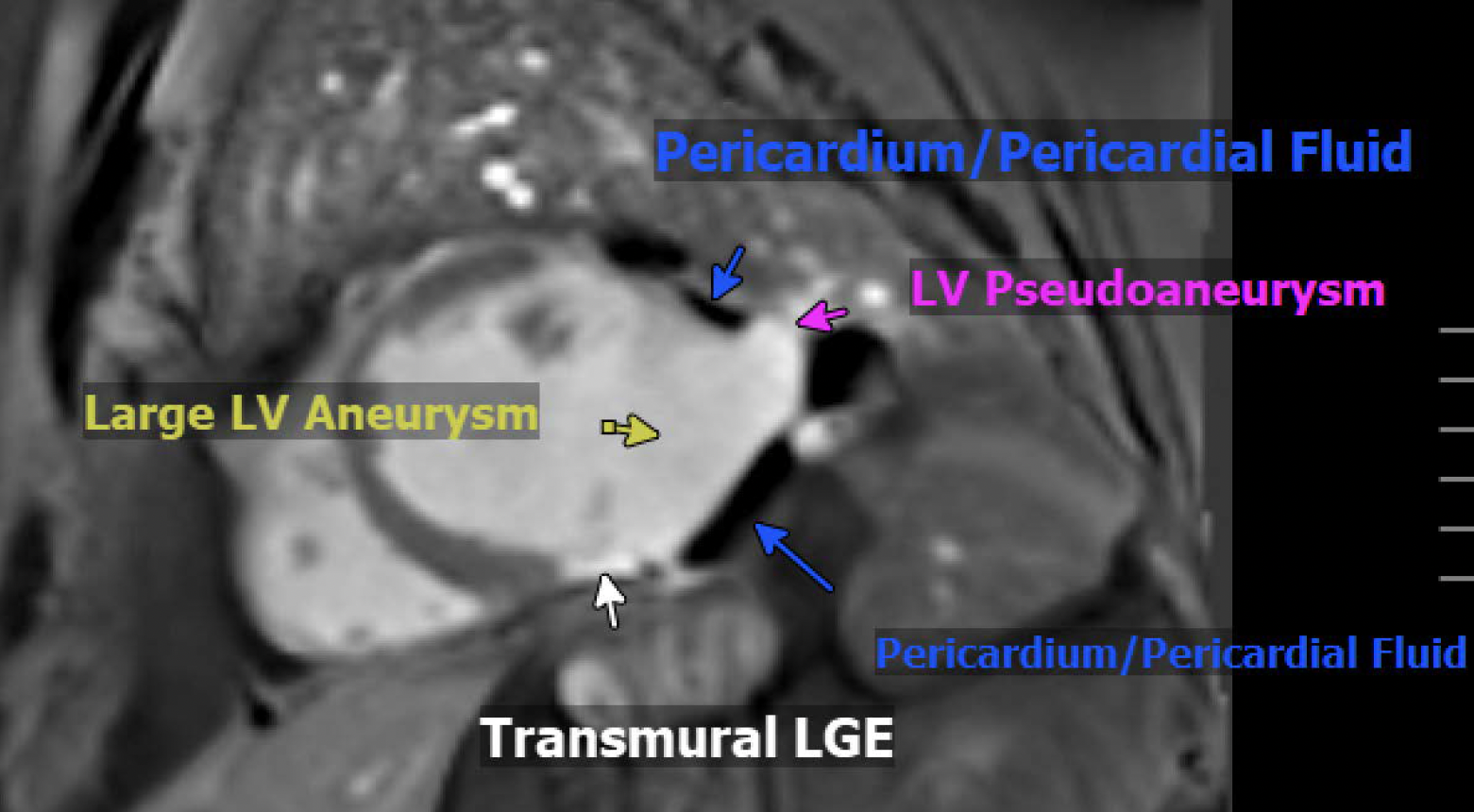 |
| Figure 6. Basal short axis PSIR at one year follow-up showing discontinuity in the myocardial signal at the rupture site, with the sac wall composed only of pericardium or fibrous tissue. |
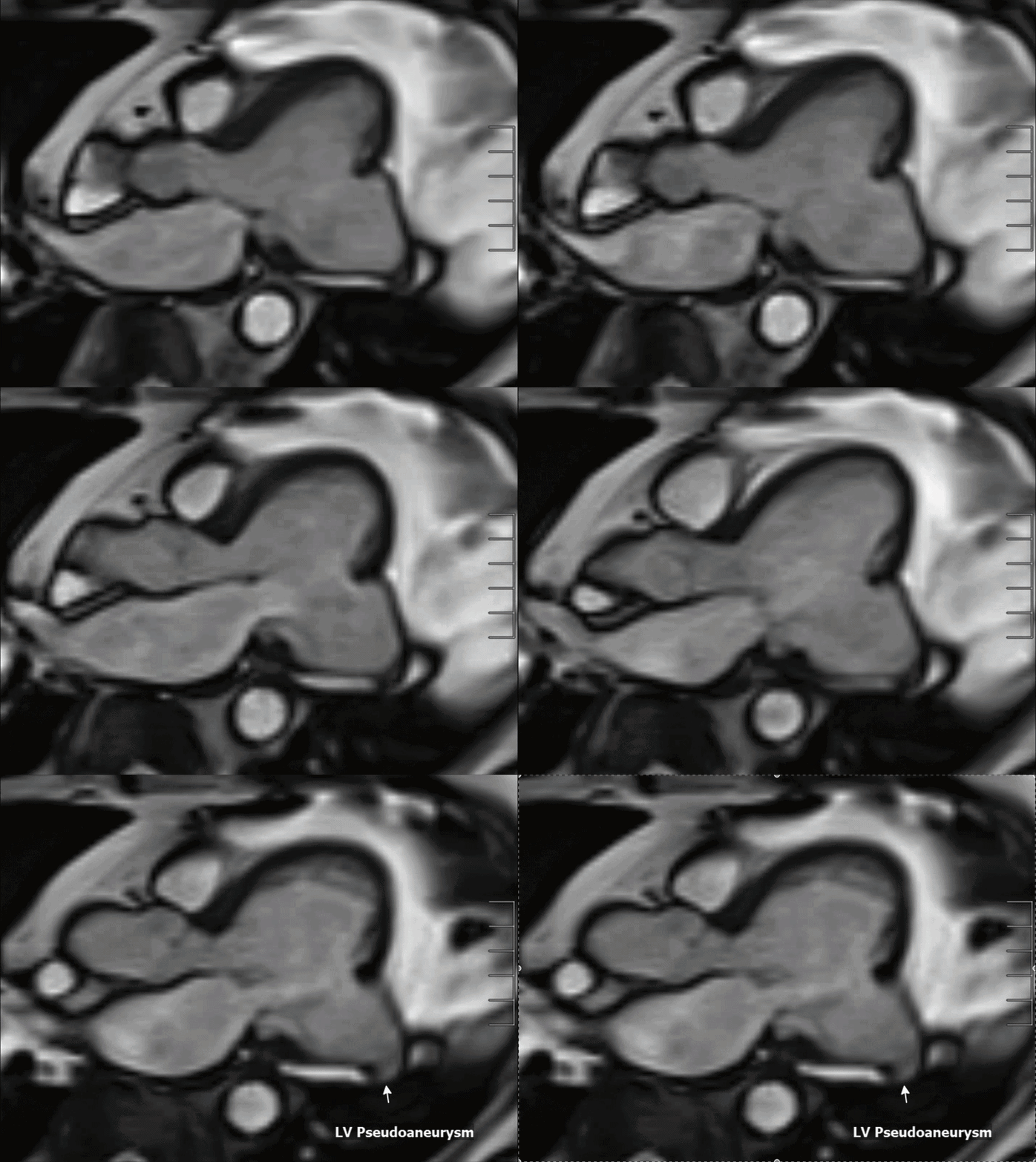 |
| Movie 1. Three-chamber cine bSSFP at one-year follow-up illustrating the large lateral wall aneurysm with a pseudoaneurysm arising anteriorly. |
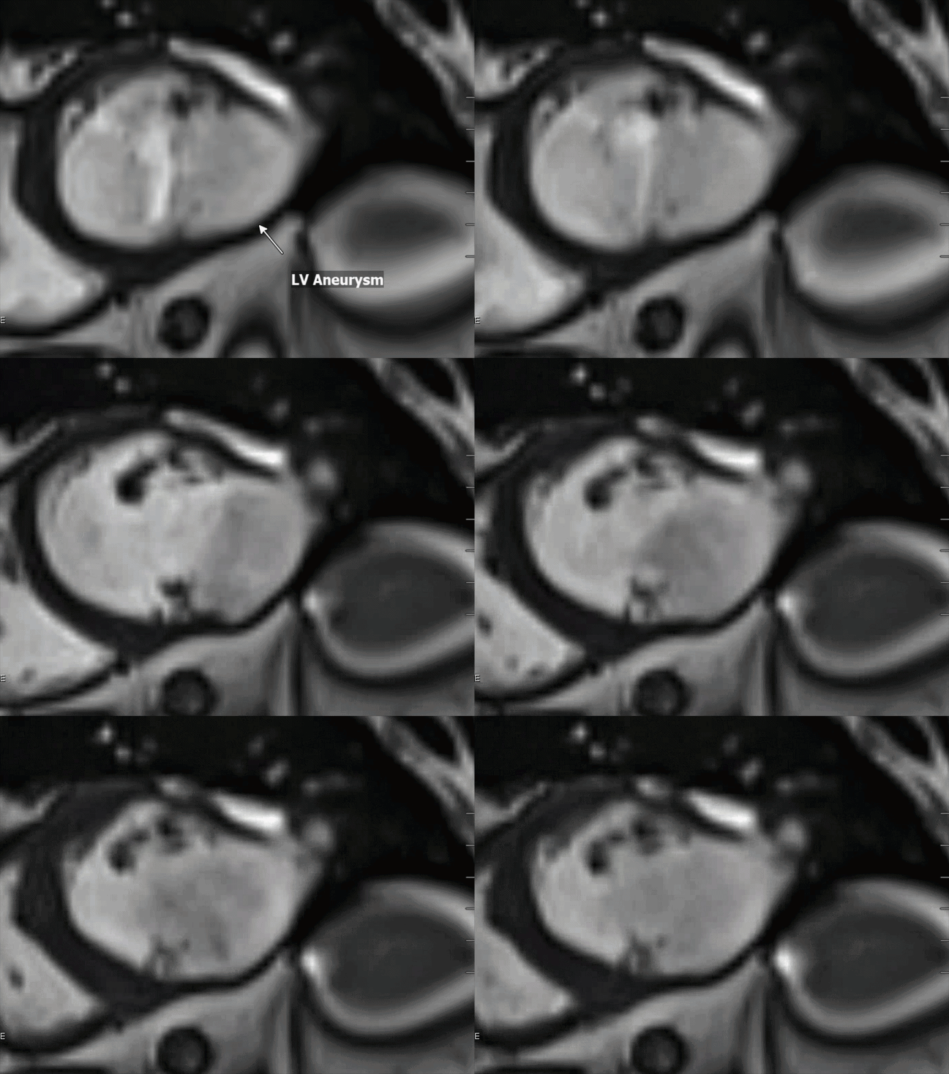 |
| Movie 2. Short-axis cine bSSFP at one-year follow-up illustrating the large lateral wall aneurysm with a pseudoaneurysm arising anteriorly. |
Conclusion
The repeat CMR was notable for the presence of a pseudoaneurysm within the LV aneurysm, that had also grown in size. This new outpouching exhibited classic features of subacute pseudoaneurysm formation such as narrow neck, absence of myocardial tissue and contained within the pericardium within the large chronic aneurysm.[1] The rupture was self-contained and led to the formation of a pseudoaneurysm. It is not definitively known whether the true aneurysm was acute or chronic due to no previous cardiac imaging. Given that the location of the aneurysm matched the infarcted coronary distribution and no prior imaging that showed a LV aneurysm, it was presumed that the aneurysm had acutely formed. Most recently, the patient was evaluated by cardiothoracic surgery. The patient was offered surgical intervention but given elevated surgical risk from age and other cardiac factors, the patient opted against surgical intervention and instead chose outpatient surveillance. The patient is planned to follow up with his cardiologist in 6 months to consider repeat imaging.
Perspective
This case illustrates the rare finding of a pseudoaneurysm arising from within an established post-infarct true LV aneurysm. The presence of both pathologies fundamentally alters clinical management. While true aneurysms can frequently be observed or managed medically, the concomitant presence of a pseudoaneurysm mandates surgical consultation due to the high risk of rupture and sudden cardiac death. The estimated risk of rupture has been reported to be between 30% and 45%.[2] A specific rupture percentage for true LV aneurysm has not been well-defined in the literature.
CMR is the gold standard for differentiating true post-infarct LV aneurysm from pseudoaneurysm and other similar pathologies such as cardiac diverticulum and congenital LV aneurysms. A true LV aneurysm typically occurs in cases of acute myocardial infarction. It is characterized by a wide neck, thinned myocardial wall later and is akinetic or dyskinetic. A pseudoaneurysm is characterized by a narrow neck and absence of myocardium.[2] Etiologies include myocardial infarction, cardiac surgery and chest trauma.[3]
A true post-infarct aneurysm also differs from a cardiac diverticulum in that a diverticulum has preserved contractility, is characterized by a narrow neck and typically is congenital in etiology.[4] Congenital LV aneurysms share some characteristic with acquired aneurysms such as akinetic/dyskinetic movement, wide neck and presence of fibrous tissue, however they differ in etiology. Congenital aneurysms result from primary cardiac dysplasia, frequent apical or submitral and often accompanied by mitral valve abnormalities.[5]
The most important distinction lies in the LGE pattern. A true aneurysm shows continuous transmural scar through a thinned but intact myocardial wall, whereas a pseudoaneurysm demonstrates a discontinuity in the myocardial signal at the rupture site, with the sac wall composed only of pericardium or fibrous tissue.[2] In this case specifically, the myocardial rupture that led to the formation of the pseudoaneurysm was most likely contained by the scar tissue of the true aneurysm that formed from the infarcted myocardium. Since a cardiac diverticulum does not have scarring, it does not demonstrate LGE uptake on CMR. Congenital aneurysms do have fibrous tissue and frequently demonstrate LGE uptake.[6]
Click here to view the entire CMR on CloudCMR.
References
- Damluji AA, van Diepen S, Katz JN, Menon V, Tamis-Holland JE, Bakitas M, et al. Mechanical Complications of Acute Myocardial Infarction: A Scientific Statement From the American Heart Association. Circulation. 2021 Jul 13;144(2).
- Lorusso R, Matteucci M, Stamatios Lerakis, Ronco D, Menicanti L, Sharma SK, et al. Postmyocardial Infarction Ventricular Aneurysm. Journal of the American College of Cardiology. 2024 May 1;83(19):1917–35.
- Konen E, Mechant N, Gutierrez C, Provost Y, Mickleborough L, Paul NS, Butany J. True versus false left ventricular aneurysm: differentiation with MR imaging – initial experience. Radiology. 2005 Jul;236(1):65-70.
- Mihali K, Harbaum L, Kreutz J, et al. Congenital left ventricular diverticulum revisited: a systematic review of clinical phenotypes, imaging features, and therapeutic strategies. BMC Cardiovascular Disorders. 2026 January; 26(165).
- Ohlow MA. Congenital left ventricular aneurysms and diverticula: definition, pathophysiology, clinical relevance and treatment. Cardiology. 2006;106(2):63-72.
- McMahon CJ, Moniotte S, Powell AJ, del Nido PJ, Geva T. Usefulness of magnetic resonance imaging evaluation of congenital left ventricular aneurysms. Am J Cardiol. 2007 Jul 15;100(2):310-5.
Case Prepared By: Robert D. Tunks, MD, MHS
Editorial Board Member, Cases of SCMR
Penn State Health, Hershey, Pennsylvania, USA