Memorial Sloan Kettering Cancer Center
Weill Cornell Medical College
Case Published in the Journal of Cardiovascular Magnetic Resonance: Click here for the link
Click here for PubMed Reference to Cite this Case
Clinical History: A 36 year old woman was admitted with pleuritic chest pain in the context of an acute COVID-19 infection. She was diagnosed with severe acute respiratory syndrome coronavirus 2 (SARS-CoV-2) by a positive reverse transcriptase PCR. Troponin-I was elevated at 13 ng/mL, and the D-dimer and other inflammatory biomarkers were elevated. She had a normal ECG with normal sinus rhythm, normal intervals, and no ST-T wave changes. Her echocardiogram showed moderately decreased left ventricular systolic function with global hypokinesis and mild right ventricular dilation and mildly decreased systolic function.
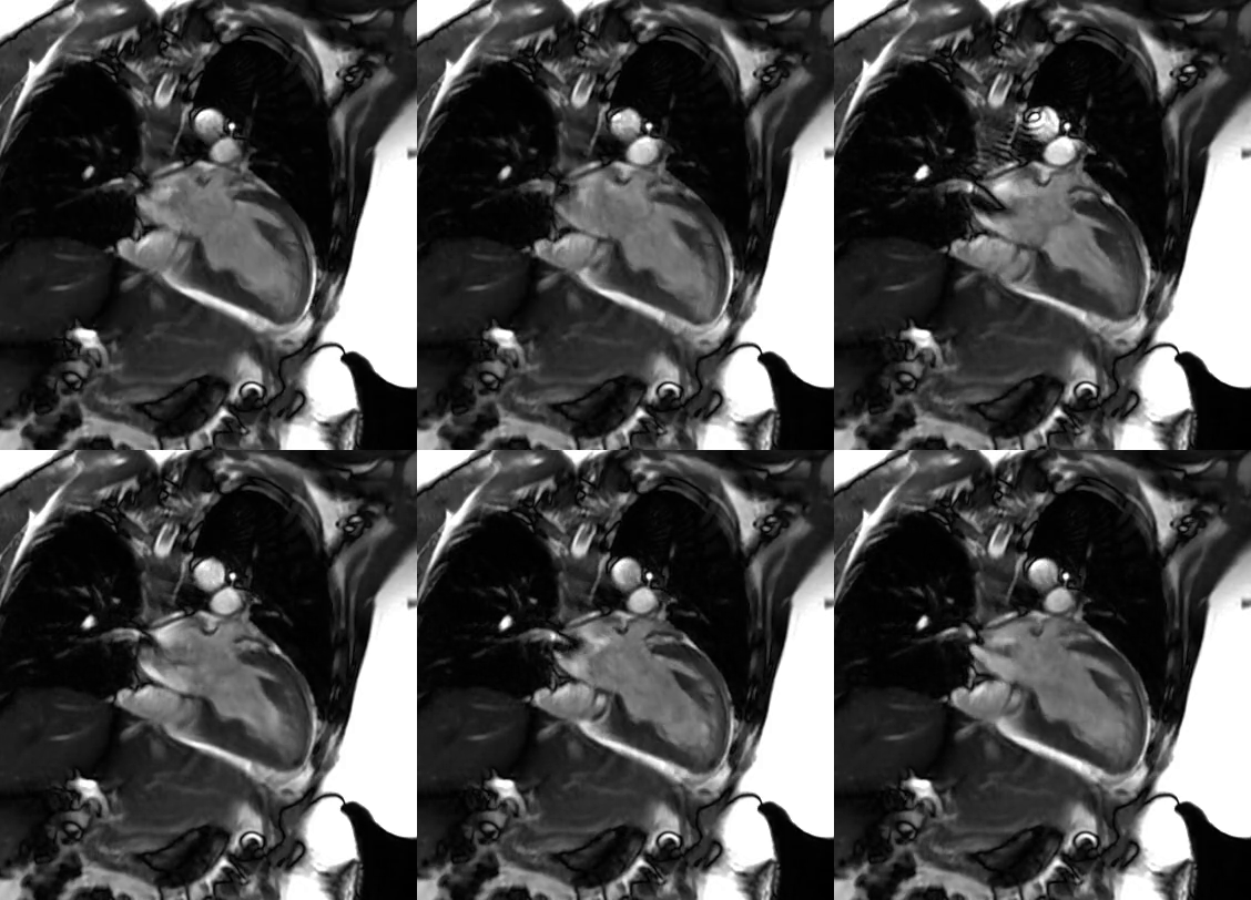
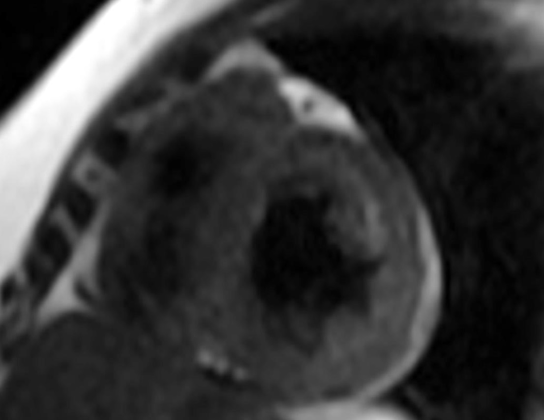
CMR Findings: Moderately reduced left ventricular systolic function (LVEF = 38%, Movie1-3) with associated elevated T2 myocardial signal (Figure 1) and epicardial late gadolinium enhancement (Figure 2). The native global myocardial T1 value was 1204 msec (3T GE, MOLLI sequence) and the global ECV was 28.4%. T1 and T2 color maps were not available.

Movie 1: Two chamber left ventricle cine SSFP with moderately decreased left ventricular systolic function.

Movie 2: Four chamber cine SSFP with moderately decreased left ventricular systolic function.

Movie 3: Three chamber left ventricle cine SSFP with moderately decreased left ventricular systolic function.

Figure 1: T2 spin echo short axis mid slice with increased signal in the lateral wall.

Figure 2: Short axis stack myocardial delayed enhancement with basal lateral wall epicardial late gadolinium enhancement (yellow arrow).
Conclusion: CMR findings were consistent with myocarditis in the setting of COVID-19 infection. She was treated with IV methylprednisolone for two days followed by a rapid prednisone taper.
Case Prepared by:
Jason N. Johnson, MD MHS
Editor, COVID-19 Case Collection
Le Bonheur Children’s Hospital, University of Tennessee Health Science Center


