


Case from: Henning Clausen, Vanessa Ferreira, Stefan Neubauer, Cameron J. Holloway
Institute: University of Oxford Centre for Clinical Magnetic Resonance Research (OCMR), John Radcliffe Hospital, Headington, Oxford, United Kingdom
Clinical history: A 69-year-old male with a history of previous myocardial infarction developed increasing exertional dyspnoea and angina four years after initial presentation. Coronary angiography revealed chronic occlusion of the right coronary artery. There was significant stenosis of the left anterior descending artery, which was successfully treated by percutaneous coronary intervention using two drug eluting stents. Left ventriculography showed a possible aneurysm in the basal inferior wall in keeping with previous infarction. There was no history of stroke during follow-up, but gradual deterioration of exercise tolerance and difficult transthoracic echocardiography windows prompted referral for cardiac magnetic resonance (CMR) ten years after initial infarction and six years after PCI.
CMR findings: CMR confirmed a massive thin walled left ventricular (LV) aneurysm measuring 5.4 x 6.4 x 4.9 cm (end diastolic volume (EDV) 125 ml) along the infero-lateral wall of the LV, almost the size of the remainder of the LV cavity (EDV of 156 ml, ESV of 53 ml) (Video 1,2). The overall systolic function was mildly impaired (LV ejection fraction 53%). On early gadolinium images there was a large adherent thrombus overlying the aneurysm, measuring 5.9 x 5.1 x 2.0 cm (Figure 1). Late gadolinium enhancement demonstrated full thickness enhancement of the aneurysm (Figure 2). Adjacent to the aneurysm, the basal inferior and infero-lateral walls demonstrated 50% subendocardial enhancement.
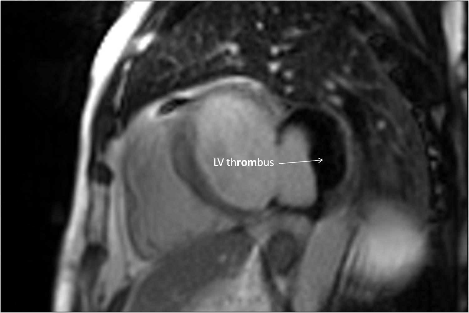
Figure 1 – Early enhancement
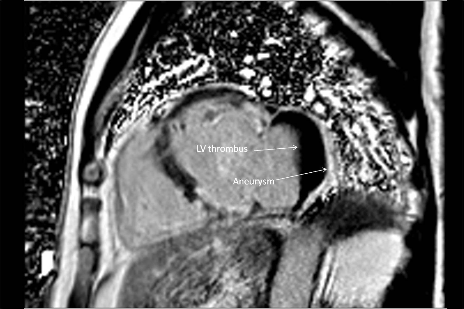
Figure 2 – Late enhancement
The patient was subsequently anti-coagulated and referred for successful aneurysmectomy. Postoperative CMR was not thought to be clinically indicated as the patient experienced marked improvement in dyspnoea over the next six months.
Conclusion: CMR provides a thorough assessment of LV aneurysms and complications,including overlying thrombi. Contrast enhanced CMR should be considered for routine assessment of LV aneurysms to assess for complications and guide surgical repair, especially when the location of the aneurysm prevents adequate ssessment by echocardiography.
Perspective: Transthoracic echocardiography has limitations when assessing LV aneurysms and identifying intracardiac thrombi compared to CMR1,2. Contrast echocardiography can improve image quality3, but CMR remains superior for detailed evaluation4 and sequential CMR assessments have been able to demonstrate short-term improvement in LV function following surgical aneurysm resection5.
References:
(1) Cheitlin MD, Armstrong WF, Aurigemma GP, et al: American College of Cardiology; American Heart Association; American Society of Echocardiography. ACC/AHA/ASE 2003 guideline update for the clinical application of echocardiography: summary article: a report of the American College of Cardiology/American Heart Association Task Force on Practice Guidelines (ACC/AHA/ASE Committee to Update the 1997 Guidelines for the Clinical Application of Echocardiography). Circulation. 2003 Sep 2;108(9):1146-62.
(2) Srichai MB, Junor C, Rodriguez LL, et al: Clinical, imaging, and pathological characteristics of left ventricular thrombus: a comparison of contrast-enhanced magnetic resonance imaging, transthoracic echocardiography, and transesophageal echocardiography with surgical or pathological validation. Am Heart J. 2006;152(1):75-84.
(3) Mansencal N, Nasr IA, Pillière R, et al: Usefulness of contrast echocardiography for assessment of left ventricular thrombus after acute myocardial infarction. Am J Cardiol. 2007;99(12):1667-70.
(4) Weinsaft JW, Kim HW, Shah DJ, et al: Detection of left ventricular thrombus by delayed-enhancement cardiovascular magnetic resonance prevalence and markers in patients with systolic dysfunction. J Am Coll Cardiol. 2008;52(2):148-57.
(5) Heatlie GJ, Mohiaddin R. Left ventricular aneurysm: comprehensive assessment of morphology, structure and thrombus using cardiovascular magnetic resonance. Clin Radiol. 2005 Jun;60(6):687-92.
Submit your case here
COTW handling editor: Kevin Steel
Have your say: What do you think? Latest posts on this topic from the forum

