


Case from: Timothy Herbst, MD; Olga Toro-Salazar, MD; Michael O’Loughlin, MD.
Institute: Hartford Hospital; Hartford, CT, USA.
Clinical history: 18 year old male with tuberous sclerosis presents for cardiac MR after an abnormal echogenic foci was identified on echocardiogram. Additional past medical history includes a giant cell astrocytoma which was resected 6 years prior.
Figure 1: Echocardiogram demonstrated a small echogenic foci at the base of the anterolateral papillary muscle in the left ventricle. Given the patient’s history of tuberous sclerosis, this finding raised the possibility of a cardiac rhabdomyoma, and the patient proceeded to have a cardiac MR for further evaluation
CMR Findings:
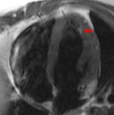
Figure 2a
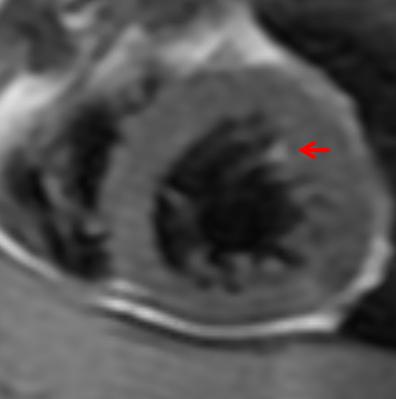
Figure 2b
Figure 2: Horizontal long axis (a) and short axis (b) double inversion recovery (IR) sequences demonstrated an approximately 7 mm focus of high signal along the base of the anterolateral papillary muscle corresponding to the location of the echogenic focus seen on echocardiogram.

Figure 3:Horizontal long axis triple IR sequence show subsequent loss of signal in this focus, compatible with fatty composition
Figure 4a
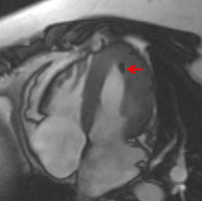
Figure 4b
Figure 4c
Figure 4d
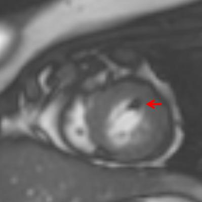
Figure 4e
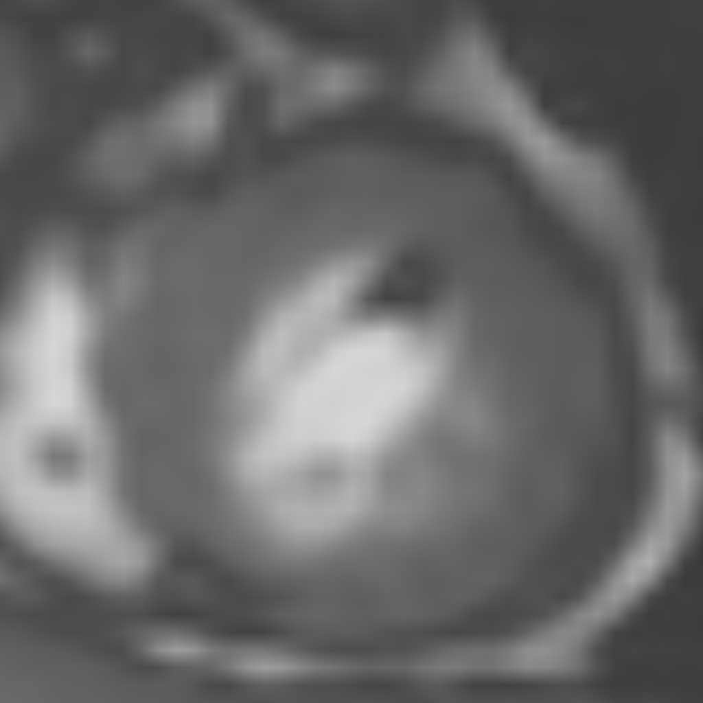
Figure 4f
Figure 4: Steady state free precession sequences in horizontal long axis (a [cine], b, c [magnified]) and short axis (d [cine], e, f [magnified]) demonstrated chemical shift artifact in this lesion with the characteristic India Ink appearance associated with the opposed phases of fat and water protons. This confirms the fatty composition of this lesion which is most consistent with a fatty focus in this patient with tuberous sclerosis. Two additional tiny hypointense foci in the lateral wall of the left ventricle on the horizontal long axis image may reflect smaller fatty deposits and are also seen to have fat signal characteristics on double and triple IR sequences.
Conclusion: In further evaluation of an echogenic focus seen at the base of the anterolateral papillary muscle on echocardiogram, cardiac MR revealed this lesion to represent focal fat in a patient with tuberous sclerosis. This lesion followed characteristics of fat on double and triple IR sequences and also displayed chemical shift artifact on the steady state free precession sequence. Myocardial focal fatty deposits have been previously described in patients with tuberous sclerosis.
Perspective: Tuberous sclerosis is an autosomal dominant inherited cutaneous syndrome characterized by the presence of multi-organ hamartomas. It has been reported that the classic diagnostic triad of mental retardation, seizures and facial angiofibromas occurs in less than half of all patients. (1) Criteria for diagnosis of tuberous sclerosis consist of both major and minor diagnostic features. (2) Since no single clinical manifestation is diagnostic for tuberous sclerosis, according to these diagnostic criteria, all clinical features should be evaluated. In patients with characteristic symptoms or skin lesions, the presence of common manifestations can allow for confirmation of the diagnosis. Alternatively, the presence of one of these findings can raise suspicion for tuberous sclerosis in undiagnosed patients who may not have outward clinical signs.
Though not the most commonly reported cardiac finding in patients with tuberous sclerosis, fatty foci in the myocardium have been previously described and characterized on CT imaging. These lesions have been identified within the interventricular septum, left ventricular wall, right ventricular wall and papillary muscles. The majority of lesions have an ovoid shape, while a minority demonstrates a more linear configuration. As seen on CT, these lesions exhibit no signs of invasive behavior and have no signs of enhancing components. A recent study by Adriaensen et al found foci of fat attenuation within the myocardium in 35 (64%) of 55 patients with tuberous sclerosis. (1) Similarly, cardiac MR examinations in patients with tuberous sclerosis have shown that these fatty foci demonstrate a well circumscribed form, location in the mid-myocardium, pure fat signal, absence of enhancement and absence of invasive behavior. (3)
On echocardiogram, these fatty foci can be visualized as areas of increased echogenicity. For this reason, these foci may be similar in appearance to a cardiac rhabdomyoma, a benign myocardial hamartoma which is a common cardiac manifestation in patients with tuberous sclerosis. Rhabdomyomas appear as solid hyperechoic masses usually located in the ventricular myocardium or ventricular septum and possible protruding into and deforming the cardiac chamber. If the lesions are multiple and small, the predominant finding is diffuse myocardial thickening which is also evident on MR imaging. On MR, rhabdomyomas manifest as a cardiac mass within or contiguous with the myocardium and can show variably increased signal intensity compared with myocardium. (5) The differing MR imaging features of rhabdomyomas and fatty foci can be helpful in the work-up of a hyperechoic myocardial focus detected on echocardiography.
Myocardial fat has been identified on CT and MR in healthy adults as well as in patients with various other myocardial diseases. Physiologic fat has been documented, primarily within the right ventricular myocardium, and the frequency and degree of physiologic fat increases with age. Myocardial fat has also been seen in the setting of healed myocardial infarction, arrhythmogenic right ventricular cardiomyopathy or dysplasia, cardiac lipoma, lipomatous hypertrophy of the interatrial septum, dilated cardiomyopathy, and cardiomyopathy with muscular dystrophy. (4)
Fatty foci in the myocardium have been shown to be present in a higher proportion of patients with tuberous sclerosis than in the general population. This suggests that the detection of fatty foci can be a helpful finding in raising the possibility of tuberous sclerosis in a patient who is yet undiagnosed, thus prompting further work-up in the correct clinical scenario.
References:
1. Adriaensen MEAPM, Schaefer-Prokop CM, Duyndam DA et al. Fatty foci in the myocardium in patients with tuberous sclerosis complex: common finding at CT. Radiology.2009; 253: 359-363.
2. Umeoka S, Koyama T, Miki Y et al. Pictorial review of tuberous sclerosis in various organs. RadioGraphics. November-December 2008. 28e32; Published online. September 4, 2008, doi: 10. 1148/rg.e32.
3. Adriaensen MEAPM, Feringa HHH, Schaefer-Profop CM et al. Focal fatty areas in the myocardium of patients with tuberous sclerosis complex: a unique finding. J Thoracic Imaging. 2011 Feb;26(1):W12-3.
4. Kimura F, Matsuo Y, Nakajima T et al. Myocardial fat at cardiac imaging: how can we differentiate pathologic from physiologic fatty infiltration. RadioGraphics.2010;30:1587-1602.
5. Grebenc ML, Rosado de Christenson ML, Burke AP et al. Primary cardiac and pericardial neoplasms: radiologic-pathologic correlation. RadioGraphics. 2000;20: 1073-1103.
COTW handling editor: M. Jay Campbell, MD

