


Case from: E. Franzì*; MC Inserra**; R.Evola*; P. Romeo**
Institute:*Department of Cardiology, **Department of Diagnostic Imaging- S.Vincenzo di Taormina Hospital–Messina, Italy
Clinical History: A fifteen years old girl with known bicuspid aortic valve was admitted with infective endocarditis due to Strept. Agalactiae and treated with antibiotic therapy. She was referred one month later to our institution for a CMR scan after a follow-up echo scan had detected a “pulsatile pouch” behind the ascending aorta.
CMR Findings: SSFP cine LVOT view and short axis view of the aortic root showed a 2.5 cm structure, pulsatile (expanding in systole and collapsing in diastole) slightly compressing the left atrium, along the intervalvular fibrous continuity (Video 1 and 2). The structure had a small neck originating at the base of the bicuspid aortic valve just below the base of the posterior cusp and was very suggestive of a pseudoaneurysm. The left ventricle was dilated. There was a severe aortic regurgitation evaluated 59 ml (regurgitant fraction 46%) on the basis of the difference between the stroke volumes of the left and the right ventricle Flow was noted through the pseudoaneurysm (Figure 1).

Video 1: Long Axis SSFP cine view of the pseudoaneurysm.
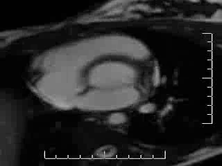
Video 2: Short Axis SSFP cine view of the pseudoaneurysm.

Figure 1: Flow long axis view: magnitude image (left) and still flow image (right) showing blood flow from the left ventricle into the pseusoaneurysm (Click to enlarge).
Conclusion: A diagnosis of pseudoaneurysm of the mitro-aortic intervalvular fibrosa was made and the patient was referred for cardiac surgery. A tissue aortic valve replacement was performed and a patch of pericardium was used to close the pseudoaneurysm.
Perspective
MAIVF is the anatomic junction between the left half of the non-coronary cusp and the adjacent third of the left cusp of the aortic valve and the anterior mitral leaflet. This area can rupture due to traumatic events or infections (1). Cardiac MRI, echocardiography (2-3) and Cardiac CT (4) can clearly visualize a mitroaortic intervalvular fibrosa pseudoaneurysm. Fatal complications can occur including compression of the coronary arteries, cardiac tamponade and mitral regurgitation, secondary to rupture of the pseudoaneurysm into the left atrium (5-6) or into the pericardial sac. We showed that CMR can be very useful for the diagnosis during functional and morphological evaluation. In addition to SSFP cines and black blood spin echo for functional (pulsatility, systo-diastolic motion) and morphology assessment, phase contrast sequences can confirm the presence of flow, further supporting the diagnosis.
References:
1) Chesler E, Korns ME, Porter GE, Reyes CN, Edwards JE. False aneurysm of the left ventricle secondary to bacterial endocarditis with perforation of the mitral-aortic intervalvular fibrosa. Circulation 1968;37:518-523.
2) Agirbasli M, Fadel BM. Pseudoaneurysm of the mitral-aortic intervalvular fibrosa: A long-term complication of infective endocarditis. Echocardiography 1999;16:253-7.
3) Yokoyama Y, Tamaki S, Kato N, Yokote J, Mutsuga M.Pseudoaneurysm from the mitral-aortic intervalvular fibrosa following endocarditis. Jpn J Thorac Cardiovasc Surg 2003;51:374-7.
4) Eduard Ghersina, Diana Litmanovicha, Yoram Agmonb, Simcha Miloc. Pseudoaneurysm of the mitral-aortic intervalvular fibrosa followingaortic valve replacement – diagnosis and dynamic evaluation withmultidetector CT and transesophageal echocardiography. Interactive CardioVascular and Thoracic Surgery 2005;4:502–504.
5) Parashara DK, Jacobs LE, Kotler MN, et al. Angina caused bsystolic compression of the left coronary artery as a result of pseudoaneurysm of the mitral-aortic intervalvular fibrosa. Am Heart J 1995;129:417-21.
6) Espinosa-Caliani JS, Montijano A, Melero JM, Montiel A. Pseudoaneurysm in the mitral-aortic intervalvular fibrosa. A cause of mitral regurgitation. Eur J Cardiothorac Surg 2000;17:757-9.
COTW handling editor: Giovanni Quarta
Have your say: What do you think? Latest posts on this topic from the forum

