Case from: Amir Dangol MD1, Mahi Ashwath MD2, Robert Gilkeson MD3, Ravi Ashwath MD1.
Institute: 1 Division of Pediatric Cardiology, University Hospital, Rainbow Babies and Children’s Hospital, Case Western Reserve University, Cleveland, Ohio, USA.
2Ohio Permanente Medical Group, Division of Cardiology, Parma, Ohio, USA.
3 Division of Radiology, University Hospital, Case Western Reserve University, Cleveland, Ohio, USA.
Clinical history: A 22 year old female was seen for a routine follow up of her apical muscular ventricular septal defect (VSD). A transthoracic echocardiogram in the apical four chamber view showed the apical muscular VSD and flow acceleration just below the moderator band with V-max of 3.7 m/s (Video 1).
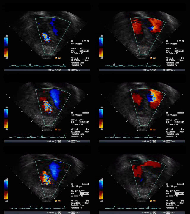
Video 1: Transthoracic echocardiogram in a 4 chamber view showing apical VSD and flow acceleration just below the moderator band raising a suspicion of a DCRV.
This raised a suspicion for the presence of a double chambered right ventricle (DCRV). A cardiac MRI was requested to further define these findings, including DCRV and also to obtain the Qp:Qs.
CMR Findings: Cine SSFP images obtained in the ventricular long and short axis (Video 2, 3) revealed a small apical muscular VSD that was contained by the muscular trabeculations in the right ventricle.
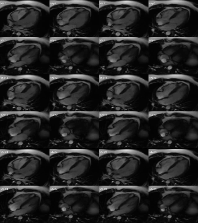
Video 2: SSFP Cine images in the 4 Chamber view showing the muscular apical ventricular septal defect contained by the muscular trabeculations in the RV.
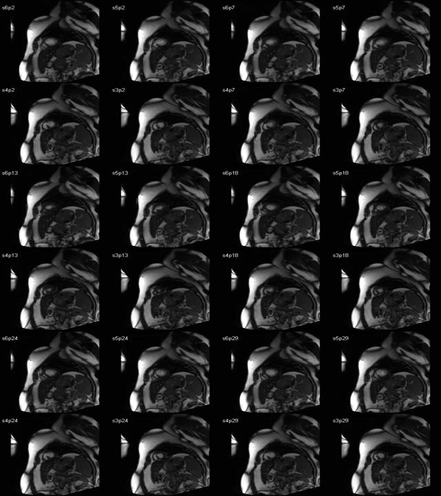
Video 3: SSFP Cine images in the short axis views showing the muscular apical ventricular septal defect.
There was a tiny restrictive communication with the right ventricular inflow apex which is a part of the right ventricular inflow and not to be confused with the right ventricular infundibular apex as described by Kumar et al [1]. The left atrium and left ventricle were normal in size. There was no RV hypertrophy. There was no evidence of a DCRV (Video 4).
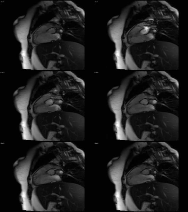
Video 4: SSFP cine images showing no evidence of a double chambered right ventricle.
First pass perfusion imaging was helpful in showing the VSD which was almost completely walled off in the right ventricular side (Video 5).
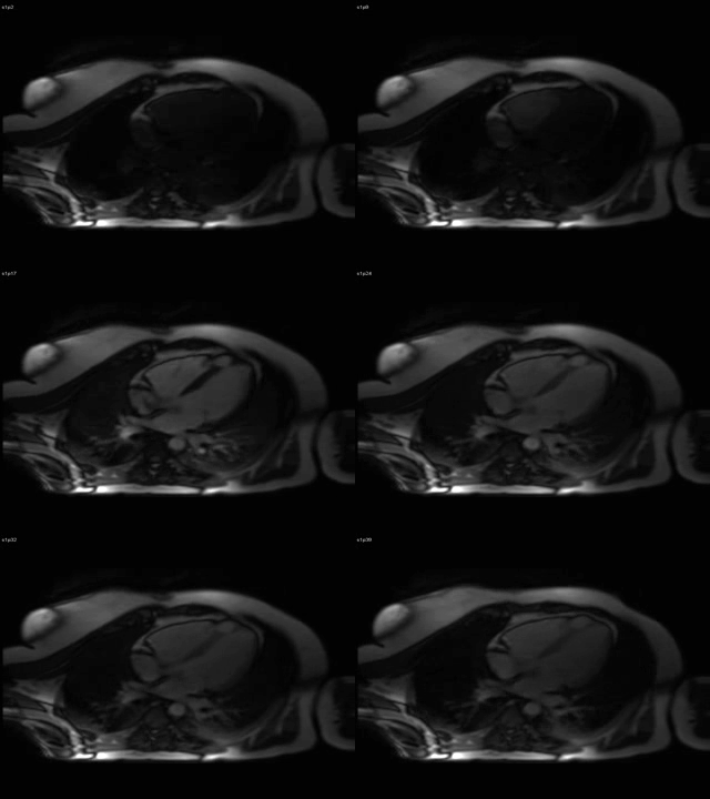
Video 5: First pass perfusion imaging in 4 chamber view showing the apical muscular VSD contained in the muscle of the RV.
Perfusion imaging showed the sequence of contrast opacification of the right side of the heart initially followed by the left side and then the VSD which was contained by the RV muscular trabeculations. It also shows mild dephasing in the RV secondary to a tiny left to right. This sequence was helpful, since it is similar to an angiogram and helped in sorting out DCRV versus a VSD. The calculated Qp:Qs was 1.1 by flow measurements across the pulmonary artery and the aorta. An in-plane phase contrast imaging would have been helpful in identifying the shunt.
Discussion and Conclusion: Although a significant proportion of muscular VSD’s are located in the apical region, little published work is available regarding the variations in its anatomy. These variations can be attributed to the alignment of the RV trabeculae that separates the apex of the right ventricular sinus (or the inflow portion) from the apex of the infundibulum. DCRV, on the other hand is an uncommon congenital heart defect in which the RV is divided into two chambers by anomalous muscle bundles. The lower pressure chamber is usually the right ventricular outflow tract (RVOT), and the higher pressure chamber is the proximal chamber which communicates with the VSD. There is a high association with VSD in about 63% to 90% of patients [2, 3]. The VSD is typically the perimembranous type but it can be located throughout the septum [4]. DCRV is a progressive disease [4], with little known about its natural history. It is therefore important to differentiate between these two entities and echocardiogram can sometimes fail to distinguish this. An accurate diagnosis is important for further management of the patient. Hemodynamically insignificant muscular VSD’s need no intervention. Hemodynamically significant apical VSD may need intervention. Transcatheter device closure of apical muscular ventricular septal defects between the left ventricle and the right ventricular infundibulum has been reported in the literature for hemodynamically significant defect [1]. So it is important to accurately define the location of the VSD and its relation to the RV inflow apex. In contrast, DCRV will most likely require surgical intervention, with muscle bundle resection.
Perspective: CMR is increasingly used for evaluating congenital heart disease. This report not only demonstrates incremental value of CMR in the assessment of complex type of muscular VSD but also highlights its importance in its ability to obtain flow data. The cine images helped us to identify its location and differentiate it from DCRV. The perfusion imaging sequence was additive and complementary in further confirming this and could prove to be a valuable sequence in some shunt lesions. The image resolution and the multiple planes that images can be obtained by CMR helped us to rule out the presence of a DCRV and understand the VSD anatomy better, which in turn helped in appropriate management and avoid a heart catheterization.
List of abbreviations:
VSD= ventricular septal defect; CMR= cardiovascular magnetic resonance imaging; DCRV= double chambered right ventricle; SSFP= steady state free precession; RVOT= right ventricular outflow tract; TTE= transthoracic echocardiogram. Qp:Qs-ratio of pulmonary blood flow to aortic blood flow.
References:
- Kumar K, Lock JE, Geva T. Apical muscular ventricular septal defects between the left ventricle and the right ventricular infundibulum. Diagnostic and interventional considerations. Circulation. 1997;95:1207-1213.
- Fellows KE, Martin EC, Rosenthal A. Angiocardiography of obstructing muscular bands of the right ventricle. AJR Am J Roentgenol 1977;128:249 –56.
- Said SM, Burkhart HM, Dearani JA, O’Leary PQ, Ammash NM, Schaff HV. Outcomes of Surgical Repair of Double-Chambered Right Ventricle. Ann Thorac Surg. 2012;1:197-200
- Alva C, Ho SY, Lincoln CR, et al. The nature of the obstructive muscular bundles in double-chambered right ventricle. J Thorac Cardiovasc Surg 1999;117:1180 –9.
COTW handling editor: Sylvia Chen, MD
Have your say: What do you think? Latest posts on this topic from the forum


