Case from: Adrian Dyer, MD
Institute: University of Texas Southwestern Medical Center, Department of Pediatrics, Division of Cardiology, Dallas, Tx
Clinical history: 15 year old boy with Tetralogy of Fallot (TOF) status post a valve-sparing repair who was referred for a cardiac MRI due to pulmonary valve insufficiency. He was a medical refugee and underwent TOF repair at 11 years old when he immigrated to the US. Per the operative report, it was a valve-sparing repair, the right ventricular pressure was half-systemic after the procedure and there was a small residual VSD by transesophageal echo (TEE). He presented to his cardiologist after being lost to follow-up and complained that he was having difficulty keeping up with his peers. His echocardiogram demonstrated free, severe pulmonary valve insufficiency, a dilated right ventricle, and a small residual ventricular septal defect. He was referred to our center for possible percutaneous pulmonary valve replacement. The cardiac interventionalist at our center requested a cardiac MRI to further evaluate his anatomy.
CMR Findings:

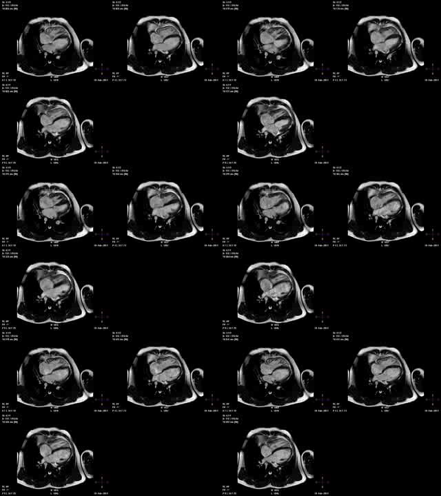
Cine 1: ECG-gated SSFP image in the ventricular long axis view (“4-chamber”) demonstrated a dilated right ventricle but also a dilated left ventricle.

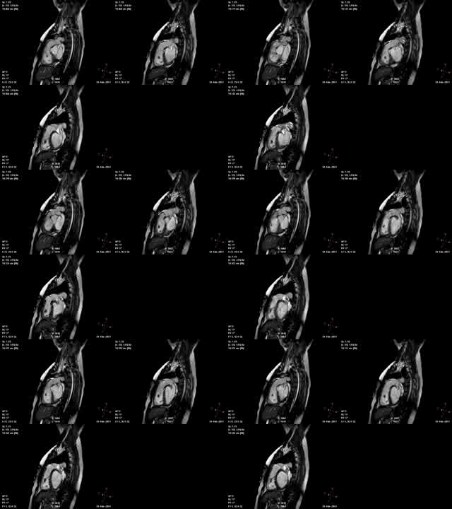
Cine 2: ECG-gated SSFP image in the right ventricular outflow tract plane (RVOT). The RVOT appeared to be native tissue with an identifiable pulmonary valve (and was confirmed with the operative note) which is currently a relative contraindication to placement of a percutaneous pulmonary valve (Melody).

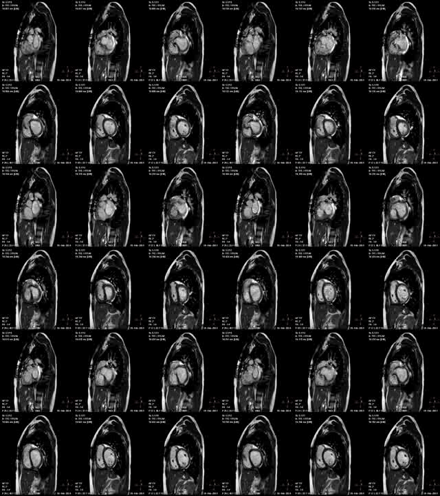
Cine 3: ECG-gated SSFP images in the ventricular short axis stack quantified biventricular dilation. There was only mild-to-moderate pulmonary insufficiency (regurgitation fraction= 17%) and estimated mild pulmonary stenosis. The residual ventricular septal defect was larger than the reported “small” by the outside echo. Of note, the VSD patch appeared to have more mobility than typically seen. The estimated Qp : Qs was 2.3 : 1.
Conclusion: .
This patient had a valve-sparing TOF repair which is currently considered the preferred mode of repair1. This involves a limited ventriculotomy with the goal of preserving the pulmonary valve apparatus even if a mild degree of stenosis remains which, over time, limits the degree of insufficiency and long-term morbidity from right ventricular dysfunction. Several papers have actually demonstrated growth of the pulmonary valve annulus after a valve-sparing repair2. This patient was interesting as he had biventricular dilation from two known complications after TOF repair: a residual VSD and pulmonary insufficiency. The VSD was quite large and seemed to be significantly contributing to his symptoms of exercise intolerance. Unfortunately due to the VSD jet being in close proximity to the tricuspid valve leaflet, an accurate VSD shunt volume could not be quantified. However, an estimated Qp : Qs was able to be calculated confirming the significance of the VSD3. In the OR after his initial repair, the residual VSD measured 2-3 mm by TEE. There is literature to support spontaneous closure of residual VSDs <2mm in size4. However, in this case, the VSD patch appeared more mobile than typical on the MRI images and had dehisced resulting in a larger defect.
Perspective:
The combination of excellent spatial resolution and flow data that CMR provides in one modality is exceedingly valuable. In this case, CMR alerted the medical team to the importance of the residual VSD and helped the patient to receive not only a pulmonary valve replacement but a much needed VSD closure.
References:
1) Apitz C, Webb GD, and Redington AN. Tetralogy of Fallot. Lancet 2009; 374:1462-71
2) Robinson JD, Rathod RH, Brown DW, et al. The evolving role of intraoperative balloon pulmonary valvuloplasty in valve-sparing repair of tetralogy of Fallot. J Thorac Cardiovasc Surg 2011; 142:1367-73.
3) Devos, DGH and Kilner, PJ. Calculations of cardiovascular shunts and regurgitation using magnetic resonance ventricular volume and aortic and pulmonary flow measurements. Eur Radiol 2010: 20:410-421.
4) Dodge-Khatami, A, et al. Spontaneous Closure of Small Residual Ventricular Septal Defects After Surgical Repair. Ann Thorac Surg 2007; 83:902-6.
COTW handling editor: Adrian Dyer, MD


