


Frank J. Raucci, Jr. MD PhD1, Jonathan H. Soslow MD MSCI1, Karla G. Christian MD2, David A. Parra MD1
Institution: Division of Pediatric Cardiology1 and Cardiothoracic Surgery2 Monroe Carell Jr. Children’s Hospital at Vanderbilt University Medical Center, Nashville, Tennessee
Clinical History
A 31 month old female was diagnosed with a large secundum atrial septal defect (ASD) following evaluation for a murmur as neonate. At that time, a separate “third chamber” was noted that appeared contiguous with the lateral left atrium that was believed to represent a cor triatriatum membrane. One pulmonary vein was seen draining into the left atrium. The other pulmonary veins were thought to drain into the “third chamber” however transthoracic echocardiography never definitively resolved this by color flow Doppler (Movie 1). She remained clinically asymptomatic although notably required three admissions for upper respiratory infections during the previous year. She was scheduled for elective closure of the ASD and resection of the cor triatriatum membrane. An intraoperative transesophageal echocardiogram was performed, which demonstrated normal pulmonary venous return and concern for a large extracardiac mass compressing the left atrium (Movie 2). The operative repair was delayed and the patient was transferred under anesthesia to the MRI suite to obtain a cardiac MRI for further evaluation of the mass prior to same day resection.
Movie 1: Transthoracic echocardiographic images: (A) Subcostal coronal view demonstrating an echogenic structure that appears to divide the left atrium and thought to represent cor triatriatum (B) Color Doppler in the same plane demonstrates a large atrial septal defect and possible entry of an upper pulmonary vein into this “third chamber”
Movie 2: Transesophageal echocardiographic images: Four chamber view with color Doppler demonstrates a large mass that does not have flow that encroaches on the left atrium causing inferior displacement of the pulmonary veins.
Advanced Imaging Findings
Cardiac MRI demonstrated normal segmental anatomy with return of the right superior vena cava, inferior vena cava, and coronary sinus to the right atrium. All four pulmonary veins returned normally to the left atrium without evidence of obstruction. A large cystic mass that measured 4 cm x 3.5 cm x 2.5 cm was located posterior to the left atrium, bordered superiorly by the main pulmonary artery, inferiorly by the roof of the left atrium, right laterally by the left atrium, and left laterally by the left upper pulmonary vein (Movie 3; presented below as separate, contiguous images positioned left-to-right). This mass had the same signal as the blood pool on SSFP imaging, but appeared to have a hyperintense signal on fat suppressed T2 sequences and medium intensity on T1 weighted sequences (Images 1A and 1B). On first pass perfusion there was no evidence of vascularization (Movie 4) and no hyperenhancement on late gadolinium enhancement imaging (Image 2).
Movie 3: Coronal cine SSFP: A large mass abutting the left atrium can be appreciated. There is no arterial compression by the mass and pulmonary venous drainage is unobstructed.
Movie 4: First pass perfusion sequence: Axial view demonstrating the mass did not enhance with contrast.
Image 2: Late gadolinium enhancement (PSIR sequences): Coronal slice through the mass shows no late enhancement
Conclusion
Following the MRI, the differential diagnosis included pericardial cyst or bronchogenic cyst [1]. It was felt that due to the very low malignant potential of the mass, surgical resection and concomitant ASD closure was prudent, so the patient returned to the operating room. A large (3.4 cm x 2.9 cm x 1.8 cm), cystic mass was resected (Image 3). Histopathologic examination demonstrated ciliated columnar epithelium with respiratory glands and the diagnosis of bronchogenic duplication cyst was made. In addition to the resection, the patient had a patch closure of the ASD without residual left-to-right shunt and trivial mitral regurgitation. She tolerated the procedure well.
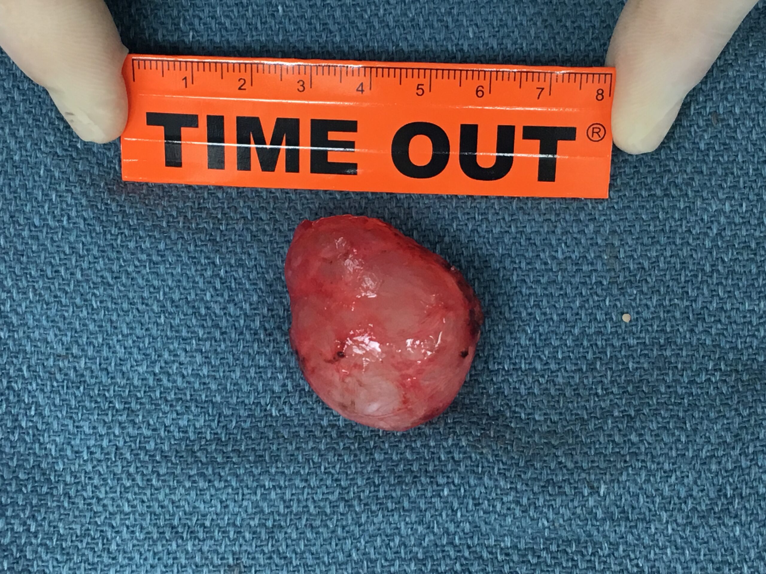
Image 3: Surgical pathology specimen of the bronchogenic cyst
Perspective
Bronchogenic cysts, also known as duplication cysts, are relatively rare with an incidence of 1 in 42,000 with approximately 70% occurring in the mediastinum [2]. After lymphoma, bronchogenic and pericardial cysts are the second most common etiologies of mediastinal cysts, each representing about one-third of diagnoses [3]. Bronchogenic cysts originate from the embryonic foregut and are comprised of a blind, fluid-filled pouch and occur most commonly at the subcarinal and right paratracheal locations [4, 5]. They are lined by ciliated epithelium and may additionally be comprised of fibrous tissue, mucous glands, cartilage, and/or smooth muscle cells [2]. Despite sharing embryonic origin with the pericardium, intrapericardial bronchogenic cysts are extremely rare with few reports in the literature [6, 7]. As in this case, MRI can be helpful in differentiating these cysts from solid tumors or vascularized structures. Most bronchogenic cysts are discovered incidentally, especially in children. Rarely, they may present with non-specific symptoms such as dyspnea, atypical chest pain, cough, or hoarseness and extremely large cysts may cause arterial, venous, or airway compression. Adults are more likely to develop symptoms or complications, such as infection or fistulization of the cyst, and there are some reports of malignant transformation [5, 8].
Cysts of the pericardium are also relatively rare, with an incidence of about 1 in 100,000, and comprise 6-7% of all mediastinal masses [3, 9]. Unlike bronchogenic cysts, pericardial cysts are thought to originate from the failure of fusion of one of the mesenchymal lacunae that form the pericardial sac and are lined with endothelium or mesothelium. Simple pericardial cysts are benign, often presenting as an incidental finding in the older asymptomatic patient. As with bronchogenic cysts, they may occasionally cause symptoms related to compression on surrounding structures and may include dyspnea, atypical chest pain, or nausea. Pericardial cysts are almost always located in the anterior mediastinum, with the majority (70%) on the right [9]. A small fraction (< 5%) are located posteriorly and finding a cystic mass in this location, as in this case, should prompt consideration of alternative diagnoses.
Click here to view all CMR images for the case on Cloud CMR
References
1. Restrepo, C.S., et al., Primary pericardial tumors. Radiographics, 2013. 33(6): p. 1613-30.
2. Limaiem, F., et al., Pulmonary and mediastinal bronchogenic cysts: a clinicopathologic study of 33 cases. Lung, 2008. 186(1): p. 55-61.
3. Lau, C.L., Davis, R.D., The Mediastinum, in Sabiston’s Textbook of Surgery, C.M. Townsend, Mattox, K.L., Evers, B.M., Beauchamp, R.D., Editor. 2004, Elsivier Health Sciences: Philadelphia, PA. p. 1738-58.
4. Castro, R., et al., Retroperitoneal Bronchogenic Cyst: MRI Findings. Case Reports in Radiology, 2013. 2013: p. 853795.
5. McAdams, H.P., et al., Bronchogenic cyst: imaging features with clinical and histopathologic correlation. Radiology, 2000. 217(2): p. 441-6.
6. Antoniou, A., et al., Intrapericardial bronchogenic cyst found incidentally during open heart surgery. Asian Cardiovasc Thorac Ann, 2012. 20(6): p. 737-8.
7. Ugurlucan, M., et al., Intrapericardial bronchogenic cyst: an unusual clinical entity. Case Rep Med, 2014. 2014: p. 651683.
8. St-Georges, R., et al., Clinical spectrum of bronchogenic cysts of the mediastinum and lung in the adult. Ann Thorac Surg, 1991. 52(1): p. 6-13.
9. Patel, J., et al., Pericardial cyst: case reports and a literature review. Echocardiography, 2004. 21(3): p. 269-72.

