


M.A.J. Becker MD, W. Yassi MD, T. Germans MD PhD
Department of Cardiology, Noordwest Ziekenhuisgroep locatie Alkmaar, Alkmaar, the Netherlands
Clinical History:
A 71-year old woman, without relevant medical history, initially presented with progressive dyspnea and orthopnea due to new onset heart failure (New York Heart Association functional class III). Her blood pressure at admission was 170/105 mmHg. A chest X-ray revealed cardiomegaly and pulmonary edema. A transthoracic echocardiogram was performed and moderately reduced left ventricular function and an echodense mass in the right atrium were observed (Image 1,2). For differentiation of various etiologies and diagnosis of the mass, cardiovascular magnetic resonance (CMR) imaging was the next step in the diagnostic strategy.
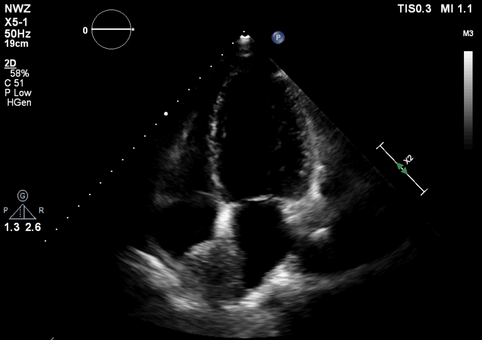

Image 1-2. Transthoracic echocardiogram apical four chamber view showing an echodense mass in the right atrium measuring 4.2 x 3.4 cm.
CMR Findings:
Cine CMR showed a mildly dilated left ventricle (end-diastolic volume 166ml (indexed 91ml/m2)) with decreased function (calculated ejection fraction 45%). Moreover, mild concentric hypertrophy was seen, possibly due to pre-existent untreated hypertension.
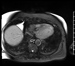
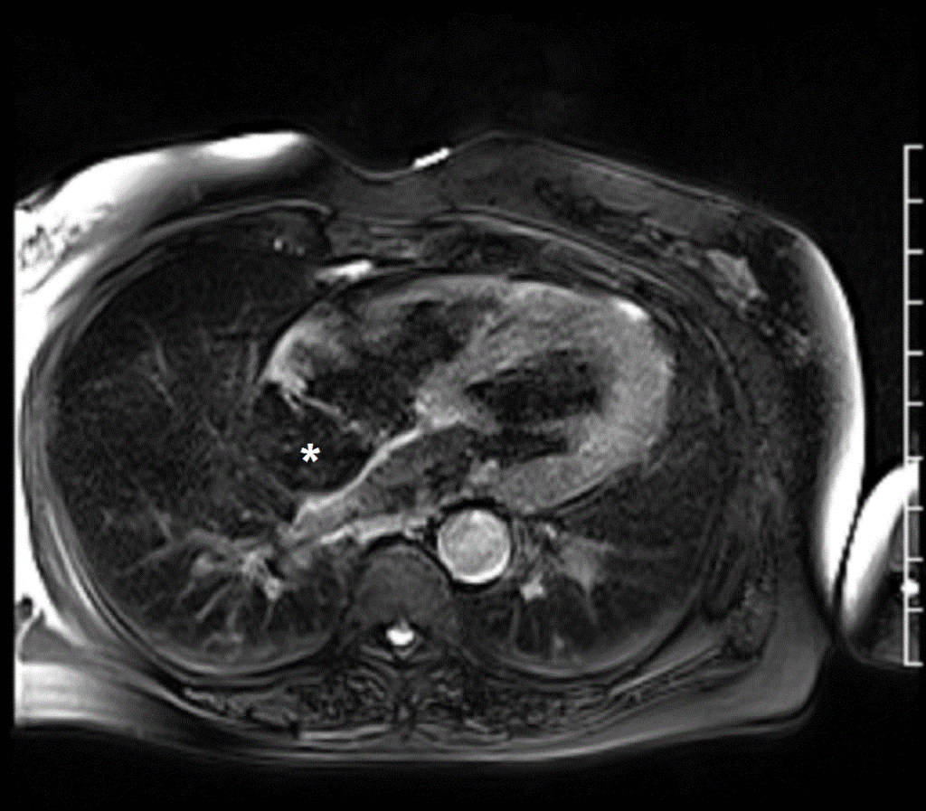
Similar to the findings on transthoracic echocardiography, a mass was found in the right atrium, localized at the right side of the interatrial septum and roof of the right atrium (Movie 1), without signs of flow obstruction on cines (Movie 2). To differentiate between etiologies, T1 and T2 weighted imaging, rest perfusion and late gadolinium enhancement (LGE) were performed. T1 weighted images demonstrated uniform hyperintensity of the mass and parts of the interatrial septum, with a signal intensity (SI) identical to the subcutaneous adipose tissue (Movie 3 and Image 3). Moreover, on fat suppressed T1 and T2 weighted black blood imaging, the right atrial mass showed complete signal suppression (Movie 4,5 and Image 4,5). Rest perfusion through the mass showed no contrast uptake, indicating a non-vascular tumor (Movie 6). After a delay of 10-15 minutes, a TI-scout was made for optimal myocardial nulling for LGE imaging. This demonstrated, as expected, the same inversion time of the mass compared to the subcutaneous adipose tissue (Image 6).
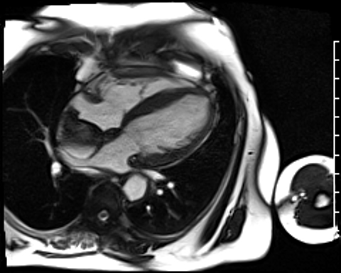
Movie 1. Cine SSFP four chamber stack outlining the extent of the right atrial mass with no evidence of obstruction of the tricuspid valve, mildly decreased left ventricular systolic function, and mild concentric left ventricular hypertrophy.

Movie 2. Cine SSFP two chamber right ventricle stack outlining the extent of the right atrial mass with no evidence of obstruction of the superior or inferior vena cavae.
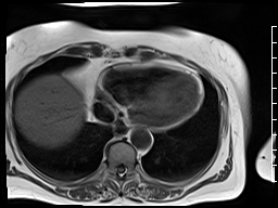

Movie 3 and Image 3. T1 weighted turbo spin echo (TSE) black blood four chamber stack illustrates uniform hyperintensity of the right atrial mass (*) and parts of the interatrial septum with a signal intensity identical to the subcutaneous adipose tissue.
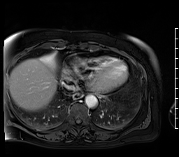

Movie 4 and Image 4. T1 weighted turbo spin echo (TSE) with fat saturation black blood four chamber stack illustrates fat suppression of the right atrial mass (white asterisk).
Movie 5 and Image 5. T2 weighted turbo spin echo (TSE) with fat saturation black blood four chamber stack illustrates fat suppression of the right atrial mass (white asterisk).
Movie 6. Four chamber rest first-pass perfusion illustrating no contrast uptake of the right atrial mass.

Image 6. Four chamber late gadolinium enhancement showing the inversion time of the right atrial mass (*) similar to the subcutaneous adipose tissue.
Conclusion:
After CMR imaging of the right atrial mass, we concluded the mass was either a right atrial lipoma, originating from the interatrial septum, or lipomatous hypertrophy of the interatrial septum, without evidence of hemodynamic obstruction or compression or infiltration of adjacent myocardial tissue. Since the patient was asymptomatic, immediate surgery was not indicated, and a watchful waiting strategy was chosen through regular outpatient department visits.
Perspective:
Of the primary myocardial tumors, which are exceedingly rare, approximately 75% are benign. Most common in adults are myxomas, typically located in the left atrium. Around 8% are lipomas, which can be located extracavitary of pericardial origin, intramyocardial, or intracavitary of subendocardial origin1–4. Lipomatous hypertrophy of the interatrial septum is more common and consists of fat deposition in the interatrial septum resulting in thickening (>2mm) of the interatrial septum, usually sparing the fossa ovalis, and is associated with higher age and obesity2,5. This results in a so called ‘dumbbell atrial septum’6.
A lipoma is characterized by a slow growth and is mainly asymptomatic, found by coincidence on cardiac imaging or post mortem5,7,8. However, depending on the size and localization, they can cause heart failure due to valvular dysfunction and flow obstruction, atrial or ventricular arrhythmias by invading the conduction system, or thromboembolic events 2,3. CMR is considered the gold standard for the non-invasive imaging and tissue characterization of cardiac masses in the heart by using T1 and T2 imaging due to its ability to differentiate between tissue characteristics9.
Click here to view case on CloudCMR.
References:
Case prepared by: Jason Johnson, MD MHS, Associate Editor, SCMR Case of the Week. Le Bonheur Children’s Hospital, University of Tennessee

