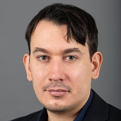
Program Chairs:

Irvin Teh, Ph.D.
Senior Research Fellow University of Leeds, UK

Kevin Moulin, Ph.D.
MRI scientist at Boston Children’s Hospital, Harvard Medical School, Boston, USA
Irvin Teh, Ph.D.
Dr. Teh is an MR physicist who leads a group focused on developing advanced diffusion MRI methods for microstructural characterization of the heart. Dr. Teh obtained his PhD in MRI Physics at Imperial College London, UK. He then took up a postdoctoral appointment at the Agency for Science Technology and Research, Singapore, and subsequently at the University of Oxford, UK. He is now a Senior Research Fellow at the University of Leeds, and the MRI Lead for the Experimental and Preclinical Imaging Centre (ePIC).
Dr. Teh’s research in cardiac diffusion MRI spans the preclinical and clinical setting, with a strong focus on the translation of demanding techniques, such as oscillating gradient spin echo, b-tensor encoding, and q-space trajectory imaging, in the heart. These cutting-edge methods offer potential new biomarkers for myocardial tissue characterization with greater specificity than existing methods. This technical development is complemented by validation using independent imaging techniques, such as synchrotron radiation imaging, validation using phantoms simulating the heart microstructure, and application in relevant patient cohorts. Dr. Teh is a keen advocate for collaborative research and co-chairs the SCMR Cardiac Diffusion Special Interest Group with Dr. Moulin.
Kevin Moulin, Ph.D.
Dr. Moulin is an MR physicist who specializes in designing and developing translational MR sequences. He obtained his Ph.D. from the University of Lyon in France under the supervision of Dr. Magalie Viallon and Pr. Pierre Croisille. He completed 5 years of post-doctoral training at UCLA and Stanford University under the supervision of Pr. Daniel Ennis. He joined Boston Children’s Hospital faculty as an Instructor in December 2022. His research activities cover the development and translational application of advanced MRI acquisitions and reconstruction techniques for chest, abdominal, and cardiovascular applications. He has worked in the emergent field of cardiac diffusion imaging in which he developed robust free-breathing and cardiac motion-compensation techniques. For this work, he was awarded an American Heart Association (AHA) postdoctoral fellowship in 2020.
Dr. Moulin’s research focuses on the development of reliable MR sequences to measure innovative quantitative biomarkers and on the translation of these sequences into robust clinical diagnostic tools. He has a special interest in cardiac diffusion imaging, motion encoding approaches, free-breathing, and cardiac motion-compensation techniques. In his latest work, he developed an approach to combine diffusion imaging and tissue displacement imaging to estimate the local strains of the cardiac cells which can help characterize the onset and progression of cardiac pathologies.
Scientific Committee:
| Dr. Irvin Teh | University of Leeds, UK | Ph.D. |
| Dr. Kevin Moulin | Boston Children’s Hospital, USA | Ph.D. |
| Prof. Frederick Epstein | University of Virginia, USA | Ph.D. |
| Dr. Sonia Nielles-Vallespin | National Heart and Lung Institute, Imperial College London and Royal Brompton Hospital, UK | Ph.D. |
| Prof. Alistair Young | King’s College London, UK | Ph.D. |
| Prof. Wu Ming-Ting | National Yang Ming University, Taiwan | M.D. |
| Prof. Alison Marsden | Stanford, USA | Ph.D. |
| Prof. Yanjie Zhu | Shenzhen Institutes of Advanced Technology, China | M.D., Ph.D. |
| Prof. Pablo Irarrázaval | Pontificia Universidad Católica de Chile | Ph.D. |
Goal
To showcase state-of-the-art techniques for imaging and modeling cardiac microstructure and motion in the heart. To bring together clinical and scientific communities and foster multi-center collaborations for advancing clinical applications and patient outcomes.
Overview
The healthy function of the heart is underpinned by the complex interactions of the microstructure and motion of the myocardium. The microstructure provides the scaffolding and connections that support the electrical conduction pathways and govern how the myocardium can deform, whilst the motion and deformation of the heart drive the cardiac output. The microstructure and motion properties of the heart are inextricably linked and can be influenced by disease processes, therapy, and remodeling.
There is considerable interest in evaluating the motion and strain properties in the heart. This includes areas such as feature tracking, tagging, strain-encoded (SENC) MRI, and phase contrast techniques e.g. tissue phase mapping and displacement encoding with stimulated echoes (DENSE). Mapping tissue microstructure is also an active field of research. We have seen the emergence of cardiac diffusion MRI as a promising tool for assessing the tissue microstructure, where the diffusion of water molecules is used as a surrogate marker for tissue microstructure. This enables quantification of tissue diffusion properties which are sensitive to cell size, integrity, and orientation, as well as microvascular perfusion. Sophisticated modeling techniques, including the use of AI, are improving image quality, facilitating the integration of electro-anatomical and hemodynamic information, and have the potential to provide patient-specific modeling of cardiac structure-function relationships.
The purpose of this workshop is to bring together clinicians and scientists to discuss the latest technological advances in the field of cardiac microstructure and motion mapping, and to identify clinical pathways for their application for the benefit of patients.
The workshop will offer cutting-edge scientific presentations, complemented by hands-on demonstrations of software packages, and a panel discussion focusing on future directions of the field. The workshop will be held in a hybrid manner to facilitate in-person and online access.
Intended audience
The intended audience for this workshop is physicists, engineers, computer scientists and physicians interested in cardiac diffusion and strain MRI and modeling.
Schedule
Venue: All sessions will be held @Blue Room, with the exception of poster sessions which will be held @Blue Pre-function Room.
Wednesday, January 29, 2025 (9:00 am – 6:00 pm)
9:00 am – 9:10 am Welcome address
9:10 am – 9:55 am Oral Session1: Exploring microstructure with cardiac diffusion imaging
10:00 am – 10:45 am Oral Session 2: Imaging myocardial dynamics with strain/motion encoding
10:45 am – 11:00 am Coffee Break (provided)
11:00 am – 11:55 am Plenary Session: State of the art of cardiac diffusion, strain and modeling.
12:00 pm – 12:45 pm Oral Session 3: Cardiac modeling, diffusion and strain analysis
12:45 pm – 1:30 pm Lunch (non-provided)
1:30 pm – 2:25 pm Oral Session 4: Clinical applications of microstructure, strain and motion encoding
2:30 pm – 3:30 pm Special Panel Session: Future Directions, Collaboration and Clinical Translation
3:30 pm – 4:00 pm Coffee Break (provided)
4:00 pm – 5:00 pm Power pitch
4:00pm – 4:15pm Oral pitch @Blue Room
4:20pm – 5:00pm Poster Session @Blue Pre-function Room
5:00 pm – 6:00 pm Poster Session
5:00pm – 5:30pm Poster Session 1 @Blue Pre-function Room
5:30pm – 6:00pm Poster Session 2 @Blue Pre-function Room
Thursday, January 30, 2025 (7:00 am – 9:00 am)
7:00 am – 7:55 am Special Educational Session: Hands-on reconstruction and analysis by academia and industry
8:00 am – 8:45 am Oral Session 5: Artificial intelligence in diffusion and strain MRI
8:50 am – 9:00 am Closing address


